 Email: tdmedical@gmail.com



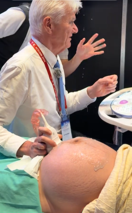
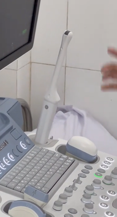
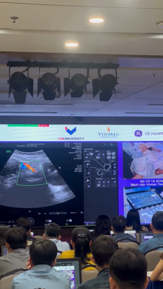
Professor Simon Meagher and Professor Ritsuko Pooh Performing Ultrasound Examinations Using the Voluson E22 System
 8.3K
8.3K
 51
51
 15-Aug-2026
 80,465

Twin-to-Twin Transfusion Syndrome (TTTS) is a serious complication that can occur in monochorionic twin pregnancies, in which vascular anastomoses within the shared placenta lead to an imbalanced exchange of blood between the two fetuses. One fetus becomes the donor twin, while the other becomes the recipient twin, resulting in complex changes in circulating blood volume, cardiovascular function, and amniotic fluid in both fetuses.
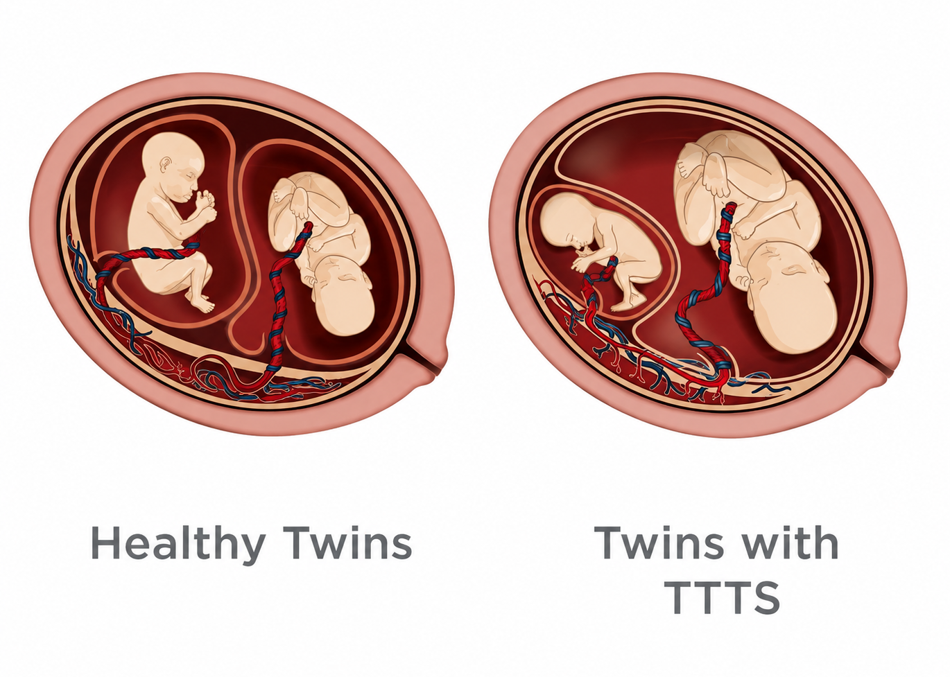
TTTS primarily occurs in monochorionic diamniotic (MCDA) twin pregnancies. In these pregnancies, the vascular systems of the two fetuses may be connected through vascular anastomoses located on or within the placenta.
The presence of these vascular connections does not necessarily mean that the pregnancy will develop TTTS. The condition arises when blood exchange through these connections becomes persistently unbalanced, causing one fetus to transfer circulating blood volume to the other.
As a result:
Donor twin → decreased circulating blood volume → reduced renal perfusion → decreased urine production → oligohydramnios.
Meanwhile:
Recipient twin → increased circulating blood volume → increased renal perfusion → increased urine production → polyhydramnios and increased cardiac workload.
This imbalance creates the characteristic sequence of changes seen in TTTS.
The most important factor is that the two fetuses share a placenta and have vascular connections between their circulatory systems.
Placental vascular anastomoses may include:
The characteristics, number, and balance of these connections influence how blood is exchanged between the two fetuses.
In TTTS, there is typically a net transfer of blood from the donor twin to the recipient twin. This is not simply a difference in fetal size but rather a hemodynamic disorder of the placental circulation.
Therefore, TTTS should not be considered synonymous with a difference in weight between the two fetuses.
The two fetuses experience different physiological consequences due to the circulatory imbalance across the placental vascular anastomoses.

In the donor twin, a portion of the circulating blood volume is transferred to the recipient twin, resulting in reduced circulating volume and decreased perfusion of the organs, particularly the kidneys. As renal blood flow decreases, urine production falls, reducing the amount of amniotic fluid and leading to oligohydramnios.
In more advanced cases, the donor twin’s bladder may become very small or difficult to visualize on ultrasound. Some fetuses may also develop growth restriction due to unequal placental sharing or associated hemodynamic disturbances.
When oligohydramnios becomes severe, the donor twin has limited space for movement and may remain closely positioned against the uterine wall. This characteristic ultrasound appearance is commonly referred to as a “stuck twin”. It is one of the findings indicating that the amniotic fluid imbalance between the two fetuses has become pronounced.
In contrast, the recipient twin receives a greater volume of blood from the shared circulation, resulting in increased circulating blood volume and renal perfusion. The kidneys respond by increasing urine production, causing an increase in amniotic fluid and resulting in polyhydramnios. This mechanism creates the characteristic polyhydramnios–oligohydramnios sequence of Twin-to-Twin Transfusion Syndrome.
However, the consequences for the recipient twin are not limited to polyhydramnios. Prolonged volume overload increases cardiac preload and forces the fetal heart to work harder. Over time, myocardial hypertrophy, diastolic dysfunction, atrioventricular valve regurgitation, or impaired cardiac function may develop. When cardiovascular compensation is no longer sufficient to manage the circulatory overload, heart failure and hydrops fetalis may occur, indicating progression to a more severe stage of the disease.
Therefore, TTTS can be dangerous for both the donor and recipient twins, although through different hemodynamic mechanisms.
Ultrasound is the primary method used to diagnose TTTS. First, the number of placentas should be determined as early as possible. This is particularly important because the risk of TTTS is associated with monochorionic twin pregnancies.

In MCDA twin pregnancies, classic TTTS is diagnosed based on an imbalance in amniotic fluid between the two sacs, with oligohydramnios in the donor twin and polyhydramnios in the recipient twin.
In addition to amniotic fluid volume, clinicians assess the fetal bladder, growth, anatomy, Doppler findings, and cardiovascular function of both fetuses.
Importantly, a difference in fetal weight is not a mandatory diagnostic criterion for TTTS.
The Quintero staging system is commonly used to describe the severity of TTTS.
| Stage | Main Characteristics |
|---|---|
| I | Polyhydramnios–oligohydramnios sequence, but the donor twin’s bladder remains visible |
| II | The donor twin’s bladder is not visible during ultrasound examination |
| III | Severe Doppler abnormalities are present |
| IV | Hydrops develops in one or both fetuses |
| V | Death of one or both fetuses |
Staging helps standardize assessment and supports pregnancy management. However, the Quintero system does not fully describe all cardiovascular functional changes, particularly those affecting the recipient twin.
Therefore, a more comprehensive assessment may require additional fetal echocardiography and Doppler ultrasound.
One of the important characteristics of TTTS is that the two fetuses must adapt to two completely different hemodynamic states.
The recipient twin generally experiences more pronounced cardiovascular effects because of prolonged circulatory volume overload.
The heart must work harder to manage the increased blood volume, which may lead to:
Some cases may develop abnormalities of the right ventricular outflow tract or pulmonary valve associated with prolonged hemodynamic changes.
Therefore, fetal echocardiography plays an important role in evaluating TTTS and provides information that cannot be fully captured by Quintero staging alone.
TTTS and selective fetal growth restriction (sFGR) can both occur in monochorionic twin pregnancies, but they are distinct conditions.
| Characteristic | TTTS | sFGR |
|---|---|---|
| Main mechanism | Imbalanced blood exchange | Unequal placental sharing |
| Key feature | Polyhydramnios–oligohydramnios sequence | Marked growth restriction in one fetus |
| Donor/recipient twins | Yes | Not necessarily |
| Umbilical artery Doppler | May be abnormal | Particularly important for classification |
| Fetal weight discordance | May be present | An important feature |
These two conditions can coexist, making assessment and management more complex.
Management depends on gestational age, disease stage, the condition of both fetuses, and the capabilities of the specialist center.
In appropriate cases of TTTS, fetoscopic laser surgery may be performed to coagulate abnormal vascular anastomoses on the placenta.
The goal of the procedure is to separate the two placental circulations, thereby addressing the underlying mechanism responsible for the imbalanced blood transfer rather than simply reducing the consequences of polyhydramnios.
Other cases may require:
Treatment decisions should be made at an experienced fetal medicine center.
Laser treatment abruptly changes the circulatory relationship between the two fetuses. Therefore, the hemodynamics of both the donor and recipient twins require time to adapt.
In the recipient twin, signs of cardiac overload may gradually improve after the abnormal blood transfer has been eliminated. Meanwhile, the donor twin may experience temporary hemodynamic changes as it adapts to the new circulatory conditions.
Doppler monitoring is therefore useful not only for evaluating TTTS but also for assessing the course of the condition following intervention.
TTTS assessment requires multiple ultrasound modalities throughout the pregnancy, including 2D imaging, fetal biometry, amniotic fluid assessment, Color Doppler, PW Doppler, and, when necessary, advanced fetal echocardiography.
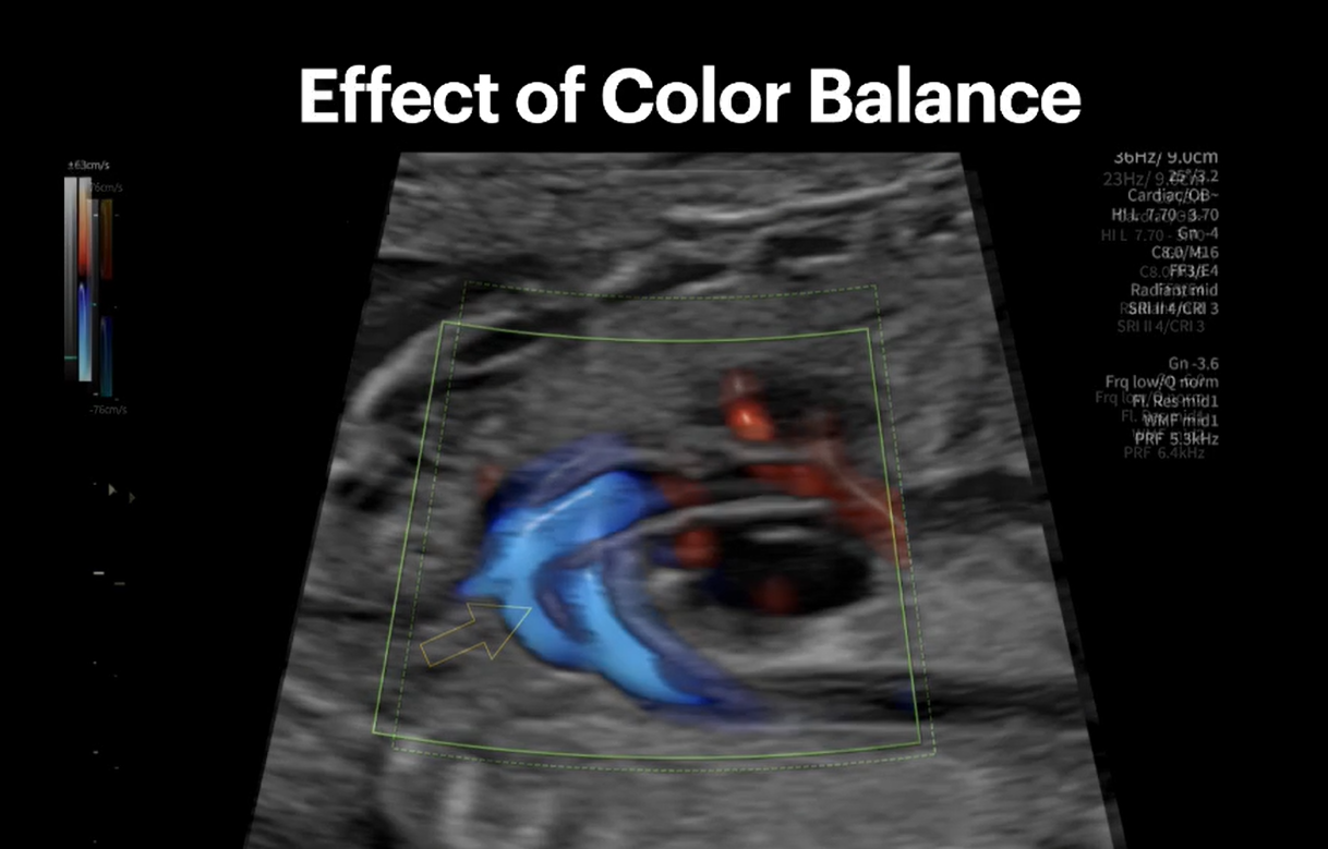
In particular, small vessels such as the ductus venosus require effective blood-flow visualization and stable Doppler waveform acquisition to minimize interference from adjacent vessels.
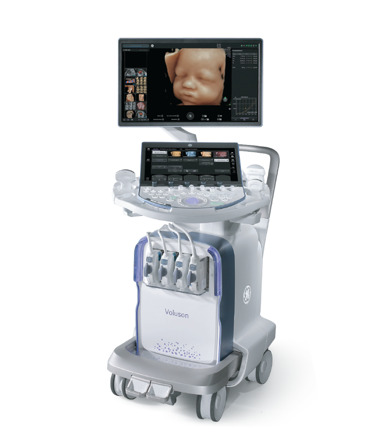
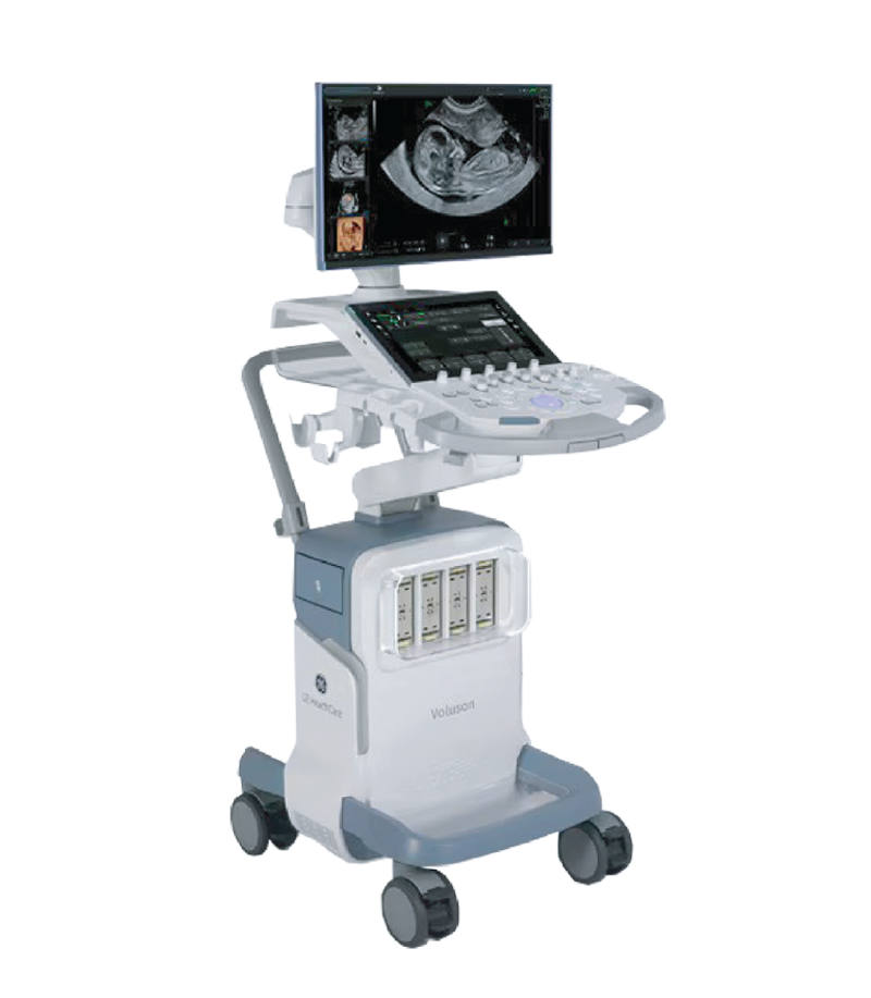
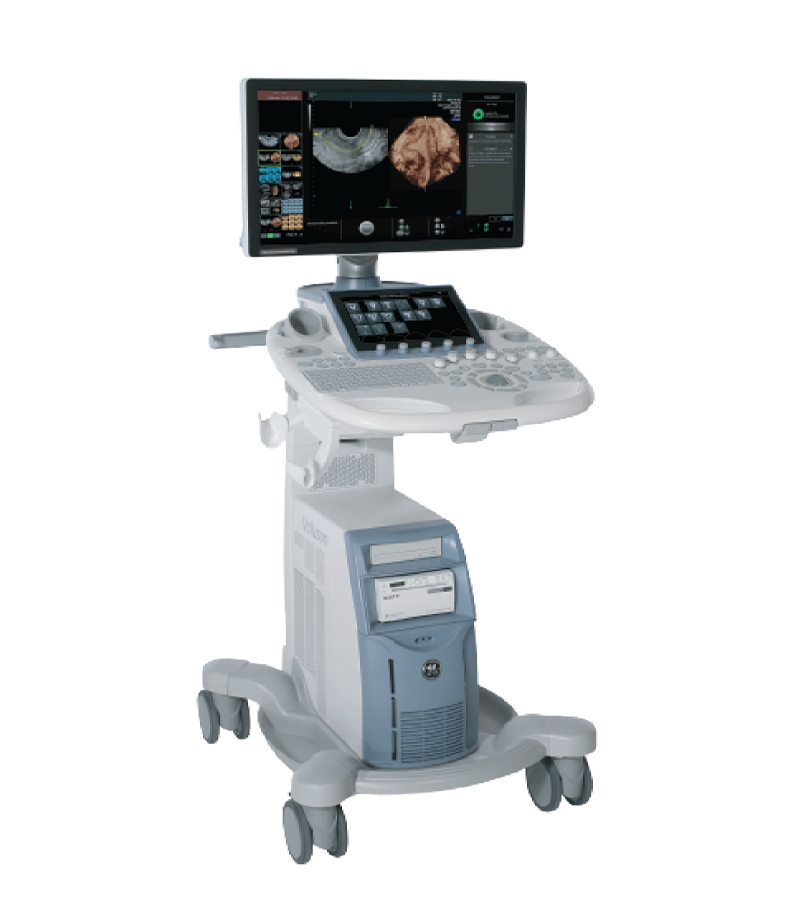
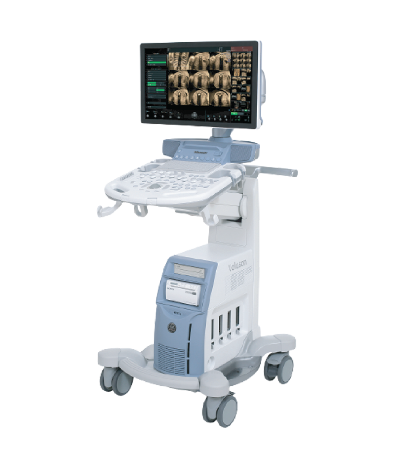
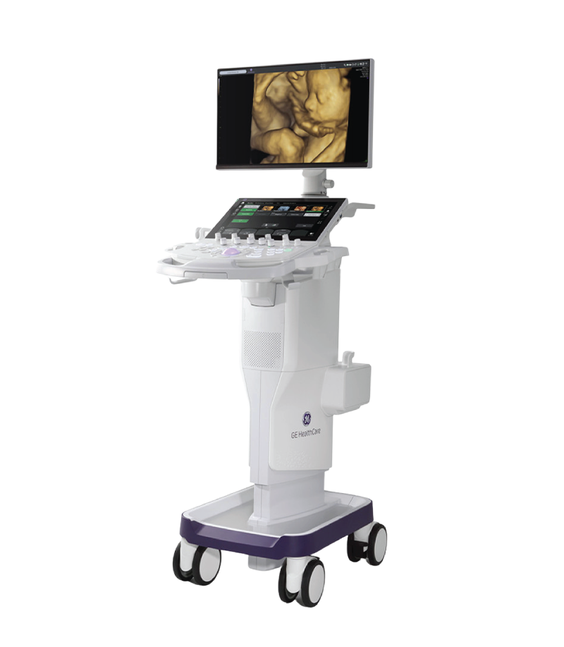
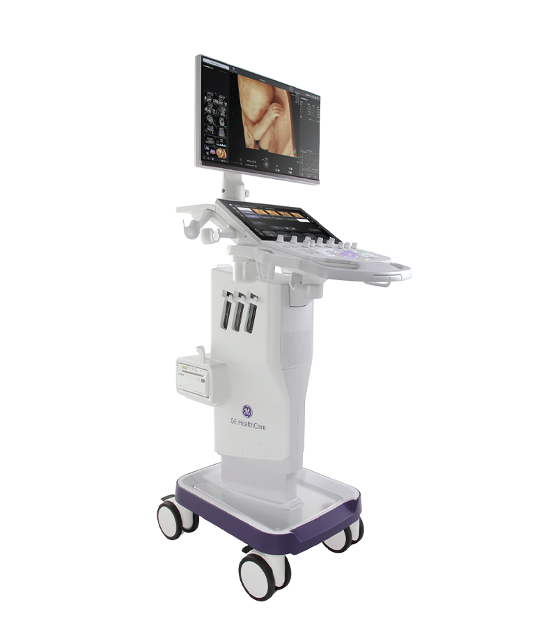
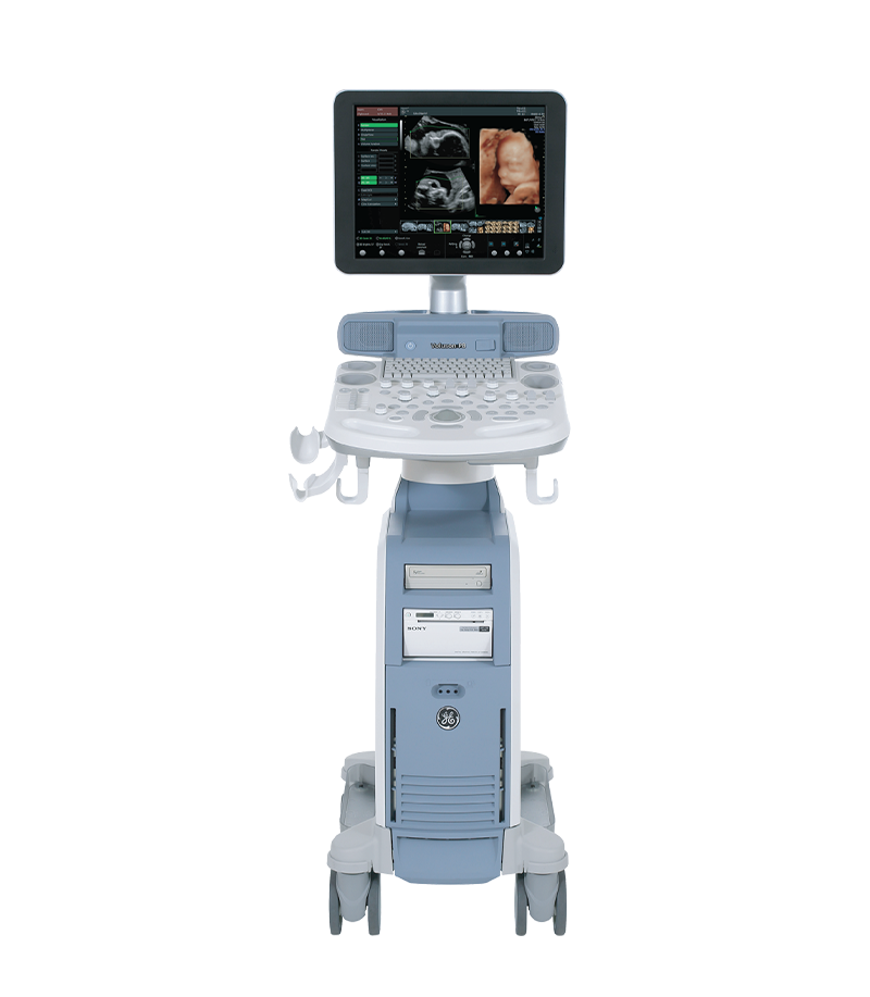
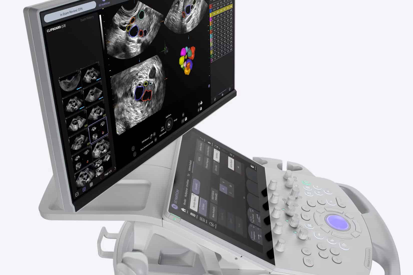
Modern obstetric and gynecologic ultrasound systems such as GE HealthCare Voluson provide a range of imaging and Doppler technologies for fetal assessment, depending on the specific system, transducers, and software packages available.
Voluson Ultrasound System
For prenatal diagnostic and fetal medicine centers, image quality, Doppler sensitivity, fetal cardiac assessment capabilities, and workflow efficiency for serial follow-up are important factors to consider when selecting an ultrasound system.
What Is a Handheld Ultrasound? A handheld ultrasound device is an ultra-compact medical imaging system, typically no larger than a smartphone or tablet, that connects directly to a smart ultrasound probe. Powered by a rechargeable...
Xem thêm
In obstetrics and gynecology, the growing need for clearer fetal visualization and more efficient examination workflows is driving the development of next-generation 3D/4D ultrasound systems. These devices are often searched for under the term “5D...
Xem thêm
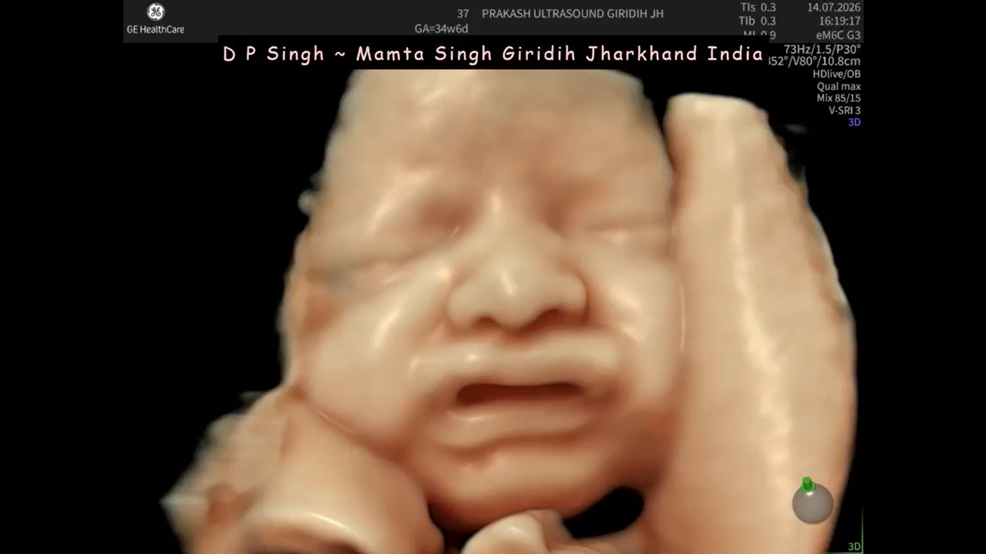
“No crying, no thinking… but definitely very expressive.” At 34–35 weeks of pregnancy, a fetus’s facial expressions can change within just a few seconds. From one ultrasound frame to the next, it can sometimes...
Xem thêm
The 9-month pregnancy journey is filled with a mix of emotions, from surprise and happiness to concerns about the baby’s development. If the first trimester is a time of adjustment and the second trimester brings...
Xem thêm
The emergence of diagnostic support software for Doppler ultrasound integrated with artificial intelligence (AI) has marked a major breakthrough, helping automate measurements, improve accuracy, and optimize workflow efficiency for physicians. What Is Doppler Ultrasound and...
Xem thêm
Congenital heart disease is among the most common congenital abnormalities in fetuses; however, prenatal detection remains challenging in routine obstetric practice. Although professional organizations such as ISUOG have issued detailed guidelines on the fetal cardiac...
Xem thêm



 Xem video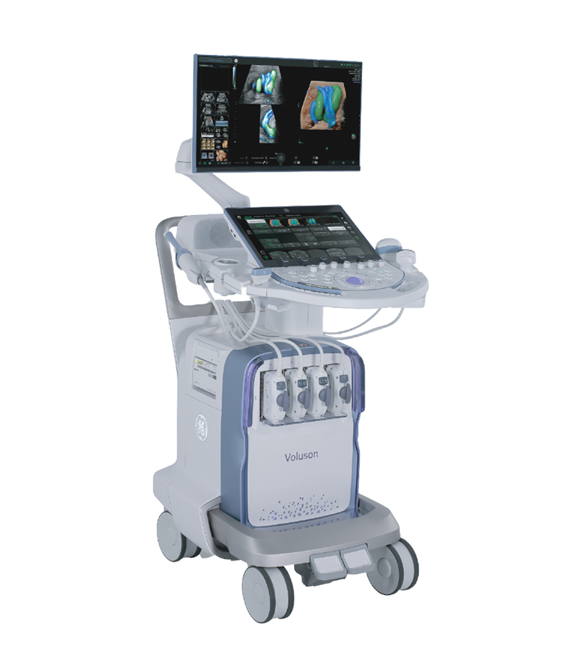




 Consult Now