 Email: tdmedical@gmail.com



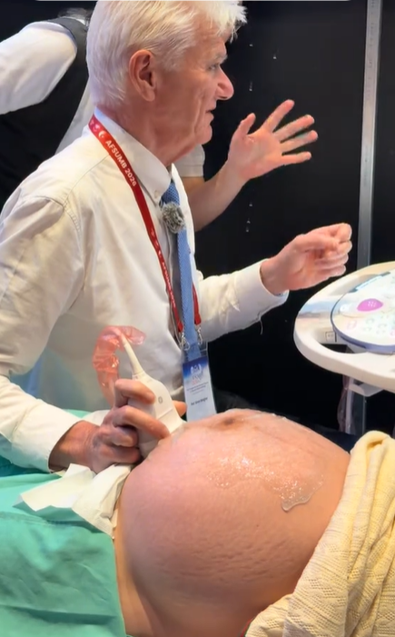
Professor Simon Meagher and Professor Ritsuko Pooh Performing Ultrasound Examinations Using the Voluson E22 System
 8.3K
8.3K
 51
51
 04-Jul-2026
 62,795

Throughout pregnancy, the moment parents see their baby’s face and movements in the womb is always one of the most precious and emotional experiences. Thanks to remarkable advances in modern medicine, 3D/4D ultrasound has become a “bridge” that allows parents to observe their baby more realistically from the very first months of pregnancy.
So, what is 3D/4D ultrasound? When is the ideal time to undergo the examination, and what benefits does this technology offer for screening fetal abnormalities? Let’s explore the details with TD TECH in the article below.
To better understand this imaging technique, it is important to first distinguish it based on the number of spatial dimensions displayed:
For advanced fetal anatomical examinations that require exceptional diagnostic accuracy, the Voluson Expert 22 and Voluson Expert 20 ultrasound systems feature a next-generation imaging architecture integrated with artificial intelligence (AI) and high-resolution rendering technology, enabling clinicians to detect subtle fetal heart and brain abnormalities at an early stage.
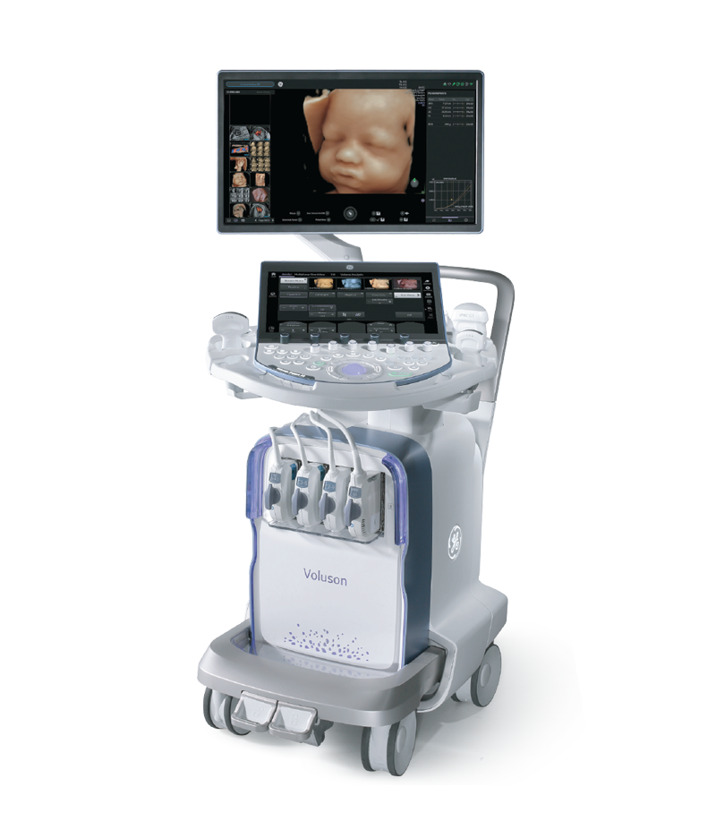
Voluson Ultrasound System
Note: Although 3D/4D ultrasound provides highly intuitive images for parents, 2D ultrasound remains the foundation for measuring fetal growth and evaluating internal organ structures. Combining these imaging modalities delivers the most accurate diagnostic results.
Beyond allowing families to see their baby, 3D/4D ultrasound plays an essential clinical role in obstetric practice.
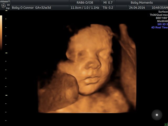
Ultrasound helps screen for and detect congenital abnormalities involving external structures, such as cleft lip, cleft palate, and hand or foot deformities (including polydactyly and clubfoot). In addition, 3D rendering technology reconstructs the fetal skull and spine, helping identify abnormalities such as spina bifida.
Watching the baby move also helps reduce maternal stress, enhances emotional bonding and happiness during pregnancy, and allows expectant mothers to prepare mentally for motherhood.
Although 3D/4D ultrasound is very safe, doctors recommend that pregnant women should not undergo the examination excessively at every stage of pregnancy. There are three important milestones to remember:
This is a mandatory screening period for assessing the risk of Down syndrome and chromosomal abnormalities. At this stage, 3D imaging helps clinicians visualize the fetus’s early anatomical development more clearly.
This is considered the ideal time for a 3D/4D ultrasound examination. By this stage, the baby’s internal organs and external features have developed significantly. The amniotic fluid volume is generally optimal, and there is still enough space in the uterus for the baby to move freely, allowing the ultrasound system to capture the clearest and most detailed images.
This stage helps detect abnormalities that may appear later in pregnancy, including those affecting the nervous system, cardiovascular system, or issues related to fetal position, the placenta, and the umbilical cord before delivery.
This is one of the most common concerns among expectant mothers, especially first-time moms. Many wonder whether watching their baby move on the screen for an extended period could affect the baby’s development.

The answer from medical experts is: 3D/4D ultrasound is completely safe for both the mother and the fetus when performed according to appropriate clinical indications. To help expectant mothers feel more confident, here are several scientific facts supporting its safety:
No ionizing X-ray radiation:
Unlike X-ray or CT imaging, ultrasound technology (including 2D, 3D, and 4D ultrasound) uses high-frequency sound waves that travel through body tissues to generate images. These sound waves do not contain ionizing radiation and therefore do not cause genetic damage or adversely affect fetal cells.
No scientific evidence of harmful effects:
After decades of widespread use in obstetric practice around the world, there is still no scientific evidence or clinical research showing that properly performed diagnostic ultrasound harms the physical or neurological development of the fetus.
Protection through modern technology:
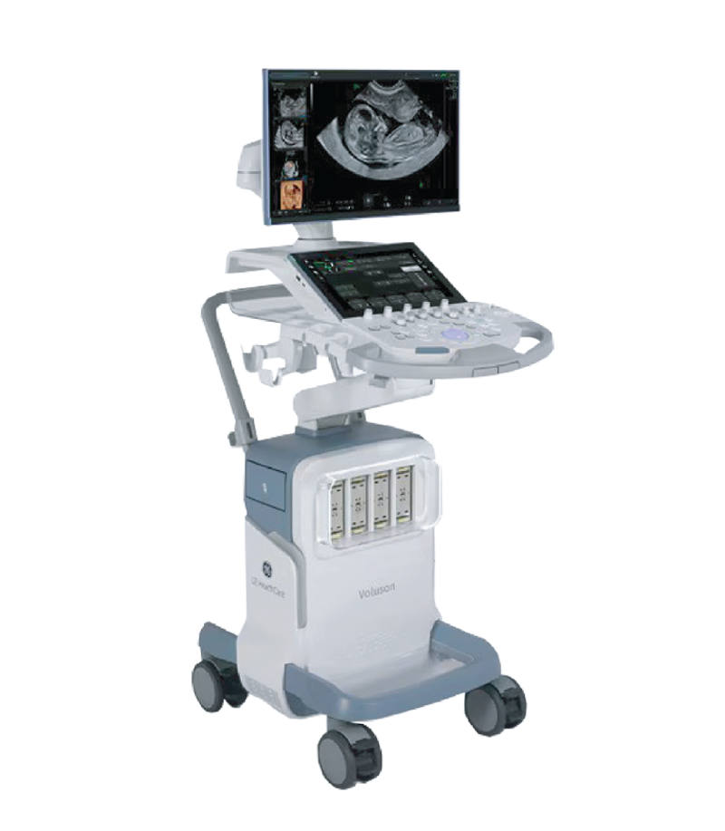
Modern ultrasound systems such as the Voluson Expert 22 and Voluson S18 are equipped with internationally recognized bio-safety indicators, including the Thermal Index (TI) and Mechanical Index (MI). These systems automatically regulate acoustic output to the lowest level required while maintaining exceptional image quality, providing maximum protection for the fetus throughout the examination.
Voluson Ultrasound System
Advice from TD TECH experts: Although 3D/4D ultrasound is safe, expectant mothers should avoid overusing this technology. Always follow your obstetrician’s examination schedule and clinical recommendations.
To ensure a smooth examination and obtain the clearest possible images, expectant mothers should keep the following tips in mind:
We hope this article has provided you with valuable knowledge about ultrasound examinations to help you prepare confidently for the journey ahead. Wishing you a healthy, happy, and wonderful pregnancy!
Every time expectant parents receive an ultrasound report, they are often confronted with a long list of unfamiliar abbreviations such as BPD, FL, CRL, NT, and AFI. To support families throughout pregnancy, TD TECH has...
Xem thêm
A 74-year-old patient was admitted to the hospital with symptoms of heart failure. Echocardiography performed using the Vivid system from GE HealthCare revealed dilated cardiomyopathy with severely reduced left ventricular (LV) systolic function, complicated by...
Xem thêm
Ultrasound is a core tool in pregnancy care, from confirming early pregnancy and monitoring fetal growth to assessing fetal anatomy, the fetal heart, and Doppler flow. Therefore, when choosing an obstetric ultrasound system, image quality...
Xem thêm
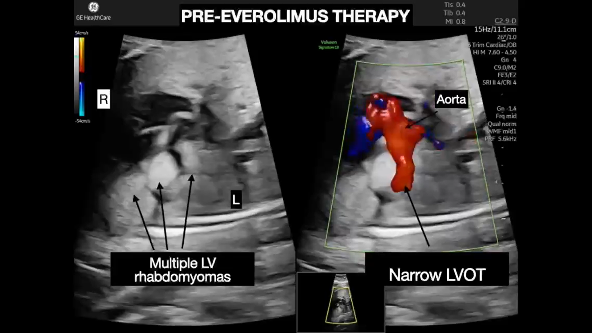
At 24 weeks of pregnancy, a routine screening ultrasound revealed multiple masses within the fetal heart. A subsequent fetal echocardiogram confirmed the presence of rhabdomyomas involving both ventricles. Although cardiac rhabdomyomas are generally benign...
Xem thêm
In obstetric practice, abnormal ductus venosus waveforms may be observed in fetal growth restriction, hydrops fetalis, arrhythmias, twin-to-twin transfusion syndrome, and certain congenital heart diseases. Therefore, ductus venosus Doppler is a valuable parameter when a...
Xem thêm
With the latest cSound platform integrated, the Vivid™ cardiovascular ultrasound systems are delivering significant value in both clinical support and diagnostic imaging. This platform represents a major step forward in image quality, workflow smoothness, and day-to-day...
Xem thêm



 Xem video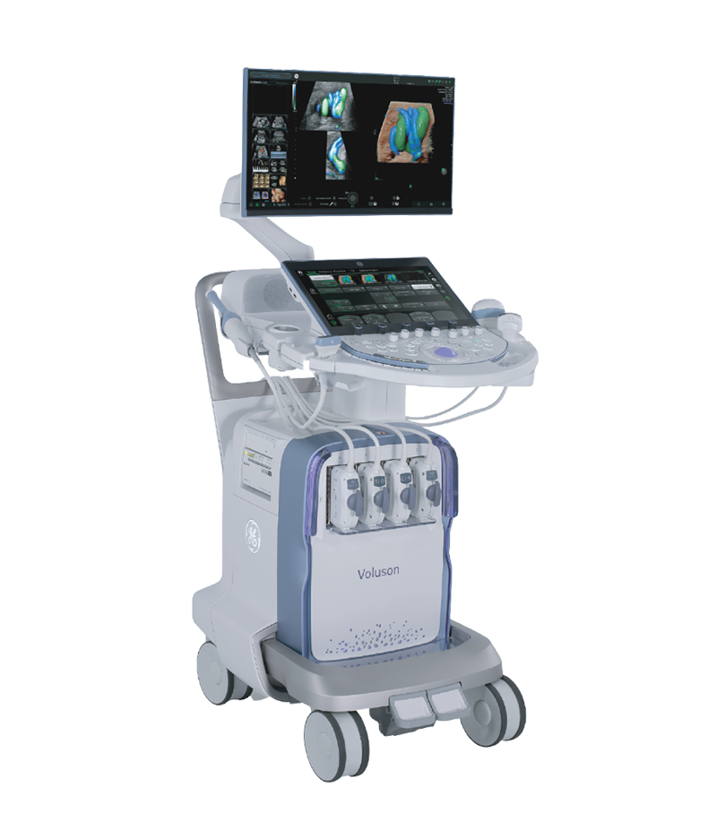




 Consult Now