 Email: tdmedical@gmail.com




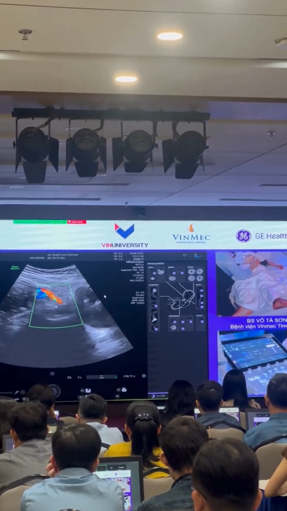
Professor Simon Meagher and Professor Ritsuko Pooh Performing Ultrasound Examinations Using the Voluson E22 System
 8.3K
8.3K
 51
51
 12-Aug-2026
 81,130

In obstetric practice, abnormal ductus venosus waveforms may be observed in fetal growth restriction, hydrops fetalis, arrhythmias, twin-to-twin transfusion syndrome, and certain congenital heart diseases. Therefore, ductus venosus Doppler is a valuable parameter when a more in-depth assessment of fetal condition is required.
The ductus venosus is a small blood vessel within the fetal circulatory system. It connects the umbilical venous system with the central venous circulation, allowing a portion of oxygen- and nutrient-rich blood from the placenta to rapidly reach the heart.
Under normal physiological conditions, most blood from the umbilical vein continues through the liver, while a portion is shunted through the ductus venosus toward the heart. This blood flow is directed toward the foramen ovale, helping deliver oxygen-rich blood to the left side of the heart and subsequently prioritizing blood supply to vital organs such as the brain and myocardium.
Because of its relatively fixed anatomical location, short length, and high blood-flow velocity, the ductus venosus is a favorable site for assessing fetal venous hemodynamics with Doppler ultrasound.
Blood flow through the ductus venosus is directly influenced by changes in atrial and ventricular pressure during different phases of the cardiac cycle.

Therefore, the DV Doppler waveform can indirectly reflect:
When cardiovascular function is compromised, these hemodynamic changes may appear in the ductus venosus waveform before certain clinical signs of decompensation become evident.
However, DV Doppler does not indicate a single specific disease. An abnormal waveform must be interpreted within the overall clinical context together with umbilical artery Doppler, middle cerebral artery Doppler, fetal cardiac assessment, fetal growth, and other ultrasound findings.
A normal ductus venosus waveform demonstrates forward flow toward the heart throughout the cardiac cycle and is generally analyzed according to the following main components:
In a normal pregnancy, flow during the a-wave remains forward. As gestation progresses, myocardial contractility and cardiac compliance improve while placental vascular resistance decreases, resulting in changes in waveform characteristics according to gestational age.
In addition to qualitative assessment, DV Doppler can also be analyzed using semi-quantitative indices. Among these, the PIV – Pulsatility Index for Veins is one of the most commonly used parameters.
The a-wave is a particularly important component when interpreting ductus venosus Doppler.
In a healthy fetus, the a-wave normally remains forward flowing. When right-sided cardiac pressure or central venous pressure increases, or when ventricular filling is impaired, a-wave velocity may decrease.
Common abnormalities of interest include:
Reduced a-wave: may reflect increased impedance to venous blood flow during atrial contraction.
Absent a-wave: blood flow approaches zero during atrial contraction.
Reversed a-wave: blood temporarily flows backward during atrial contraction and is considered an important hemodynamic finding.
An absent or reversed a-wave does not correspond to a specific diagnosis on its own, but it may suggest impaired cardiovascular function or indicate that the fetus is approaching a stage of circulatory decompensation in certain conditions.
Proper technique is essential for obtaining a reliable waveform.
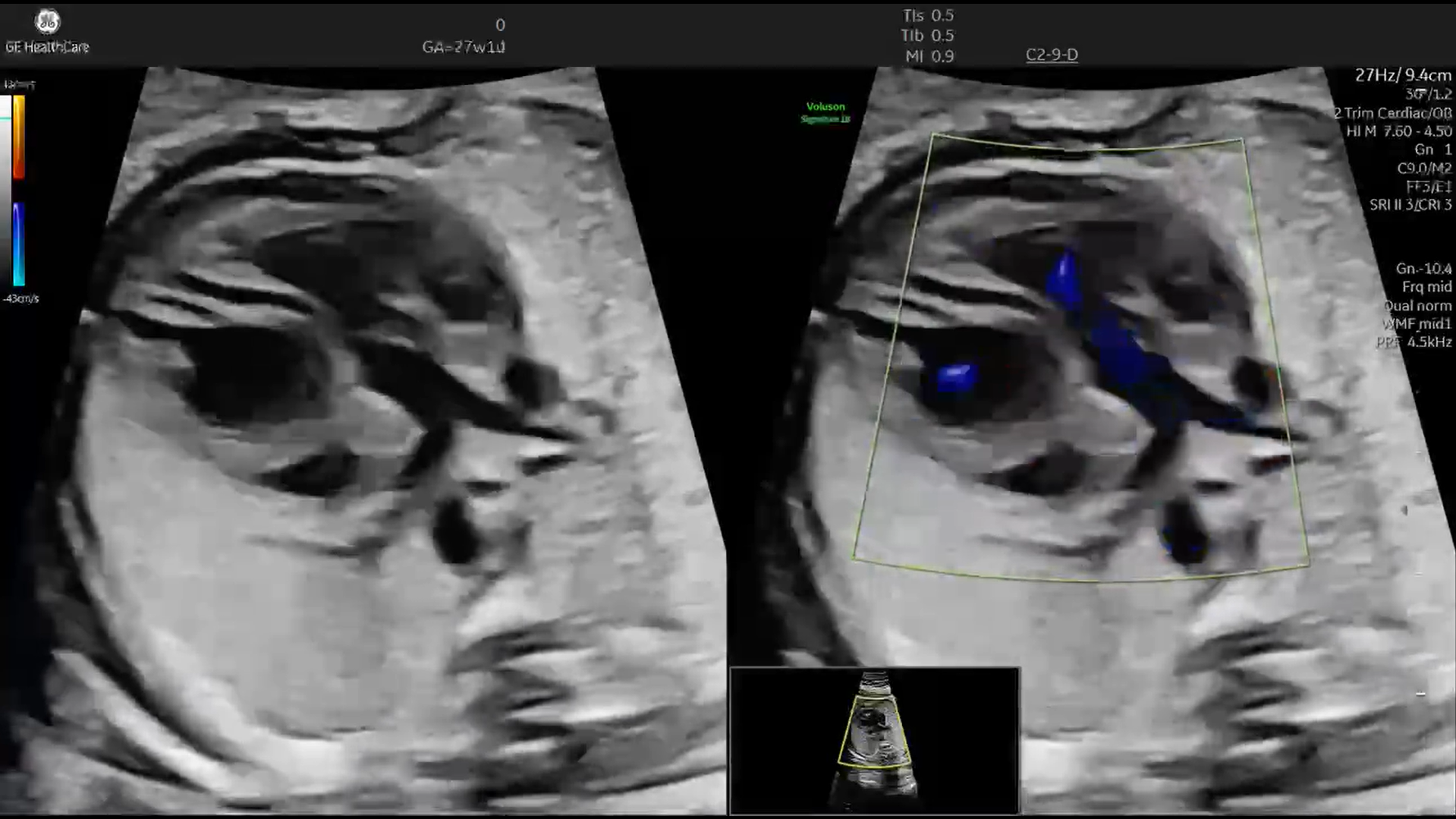
The ductus venosus is usually identified using color Doppler in a midsagittal view of the fetal abdomen or an appropriate transverse plane. Because blood-flow velocity at the ductus venosus isthmus is higher than in adjacent veins, color Doppler helps distinguish the vessel more clearly.
When pulsed-wave Doppler is used, the sample volume should be positioned at the narrow isthmic portion near the origin of the DV from the umbilical vein. A small sample volume, generally around 2 mm or less, helps minimize contamination from adjacent vascular signals.
Several factors should be optimized:
If sampling is inaccurate, signals from the umbilical vein or hepatic veins may distort the waveform.
One of the most important applications of ductus venosus Doppler in obstetrics is the monitoring of fetal growth restriction, particularly early-onset FGR.
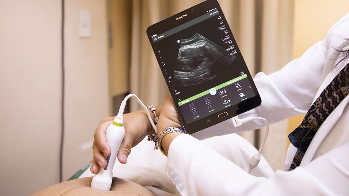
In fetuses with FGR caused by placental insufficiency, hemodynamic deterioration often begins with increased resistance in the placental circulation. Umbilical artery Doppler may show an elevated PI, followed by progression to absent or reversed end-diastolic flow.
The fetus may subsequently redistribute its circulation in an attempt to protect the brain, which can be reflected by changes in middle cerebral artery Doppler. As hypoxia becomes more severe and begins to compromise cardiac function, abnormalities may start to appear in the ductus venosus Doppler waveform.
A reduced, absent, or reversed a-wave in the context of early-onset FGR may indicate later-stage cardiovascular compromise and can be valuable for assessing the risk of fetal decompensation.
Hydrops fetalis may occur in association with various conditions, including cardiovascular abnormalities, fetal anemia, chromosomal disorders, infections, arrhythmias, or complications of twin pregnancy.
Many of these conditions can increase central venous pressure or impair cardiac function and may therefore alter the ductus venosus waveform.
When hydrops fetalis is detected, DV Doppler may help assess the extent of circulatory compromise. An abnormal a-wave in the presence of hydrops is generally an indication for more detailed cardiovascular evaluation.
In fetuses with supraventricular tachycardia (SVT), an excessively rapid heart rate can shorten ventricular filling time, reduce the efficiency of cardiac contraction, and increase central venous pressure.
If the condition persists, the fetus may develop heart failure and hydrops fetalis.
Ductus venosus Doppler can reflect these changes through alterations in waveform morphology and reduced forward flow during different phases of the cardiac cycle. Therefore, this technique may be incorporated into the hemodynamic assessment and monitoring of treatment response in fetal arrhythmias.
In monochorionic twin pregnancies, twin-to-twin transfusion syndrome (TTTS) causes an imbalance in circulation between the donor and recipient twins.
The recipient twin typically experiences increased circulating blood volume and increased cardiac workload, while the donor twin may develop hypovolemia and increased placental vascular resistance.
Ductus venosus Doppler plays a role in assessing the degree of hemodynamic compromise, particularly when an absent a-wave, reversed a-wave, or abnormally elevated PIV is present.
Severe venous Doppler abnormalities form part of the assessment of TTTS severity. Following laser treatment of placental vascular anastomoses, DV Doppler may also be monitored to evaluate circulatory adaptation in both fetuses.
Congenital heart disease can alter venous blood flow as a result of structural abnormalities, increased intracardiac pressure, or impaired myocardial contractility.
In certain obstructive right-sided cardiac lesions, such as abnormalities of the tricuspid valve or pulmonary valve, the likelihood of a reversed a-wave may increase.
During the first trimester, an abnormal DV Doppler waveform combined with other markers such as increased nuchal translucency may contribute to risk stratification and may be one of the factors prompting clinicians to consider more detailed fetal cardiac assessment.
However, DV Doppler cannot replace fetal echocardiography. When an abnormal waveform is detected, comprehensive evaluation of cardiac structure and function is necessary to determine the underlying cause.
Modern obstetric ultrasound systems increasingly incorporate highly sensitive Doppler technologies, advanced signal processing, and automated measurement tools. These capabilities are particularly important when examining small vessels such as the ductus venosus, where accurate sample-volume placement and stable waveform acquisition directly influence data quality.
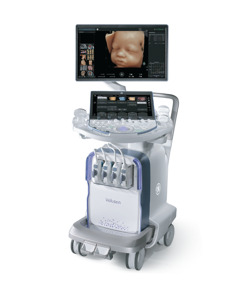
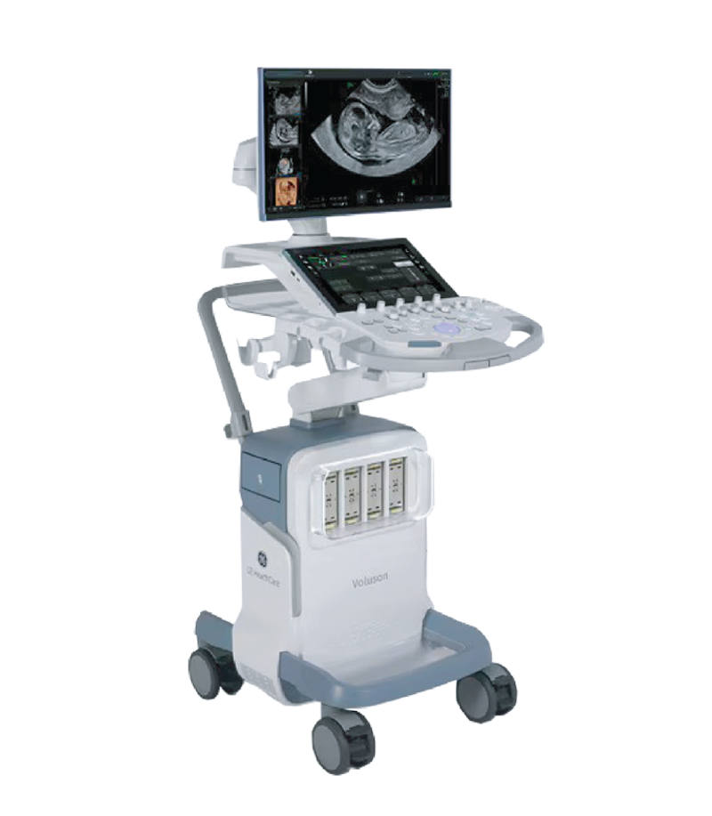
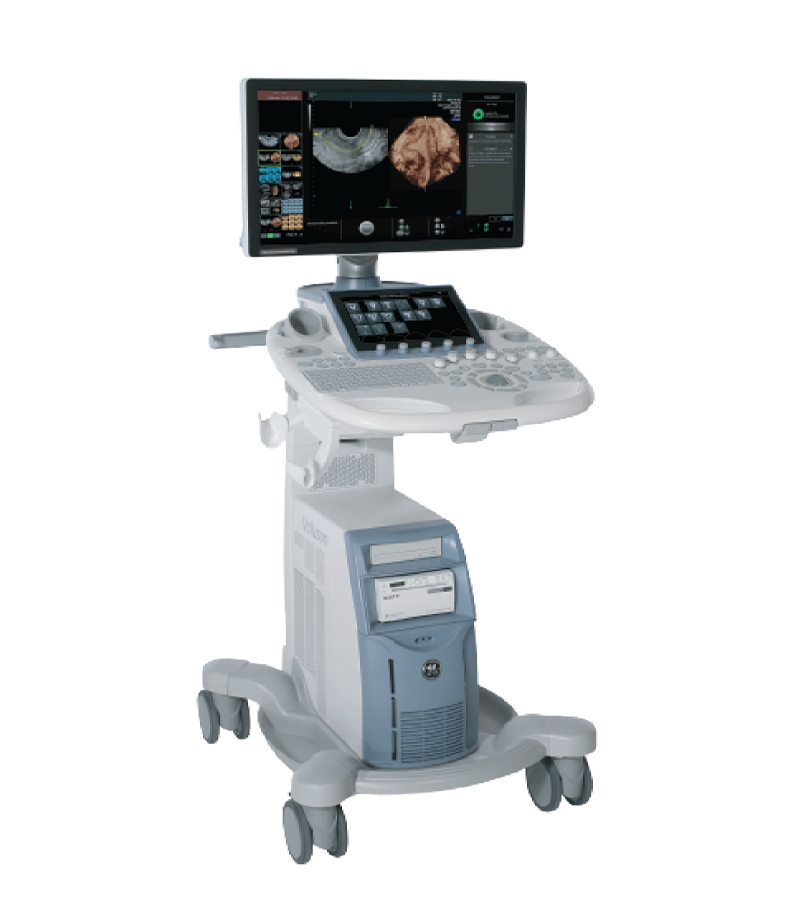
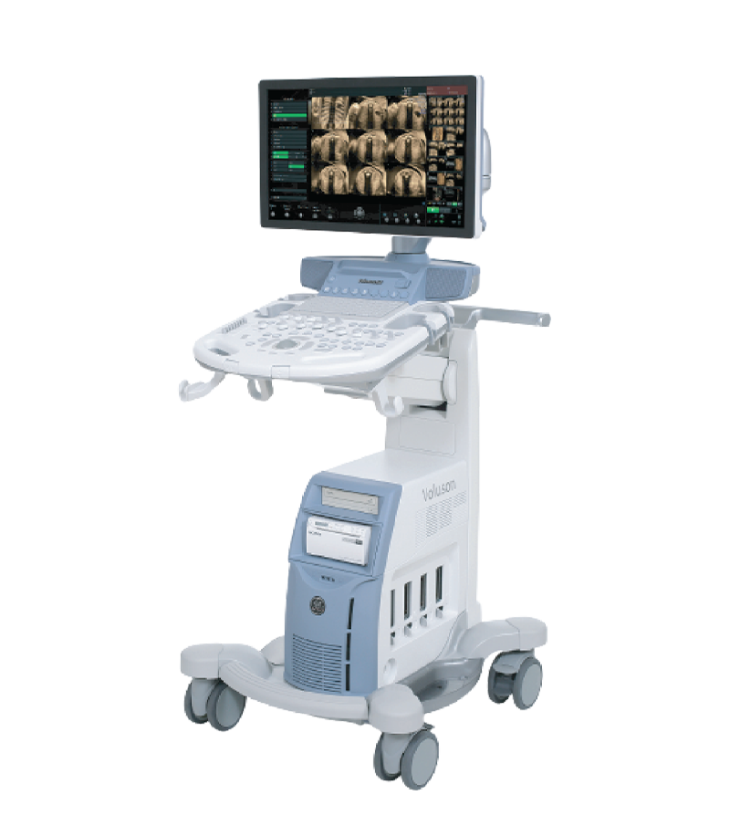
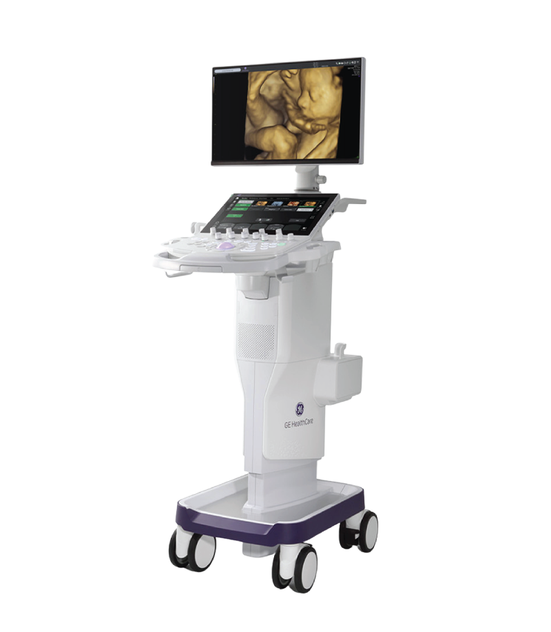
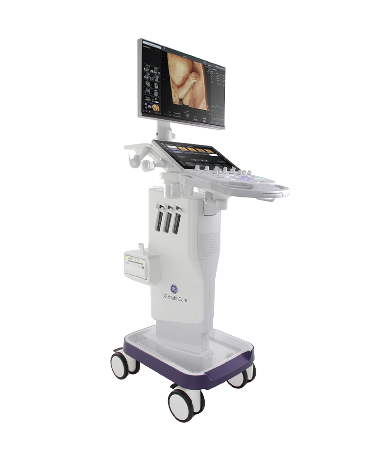
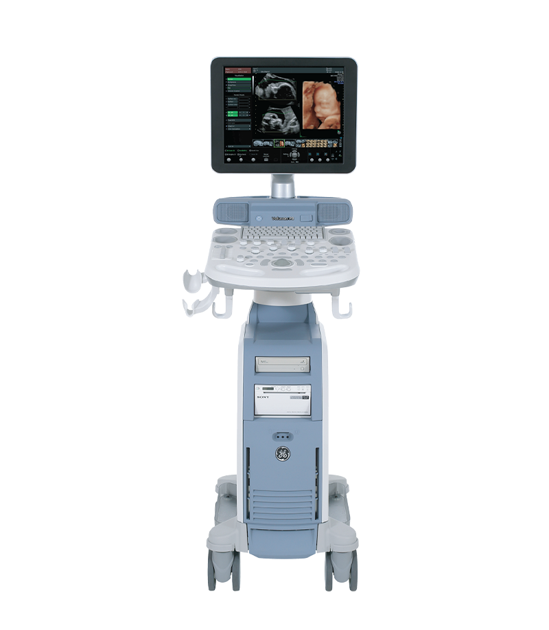
Obstetric and gynecologic ultrasound systems such as the Voluson series from GE HealthCare can provide a range of technologies supporting Doppler imaging, 2D imaging, 3D/4D imaging, and advanced fetal assessment, depending on the system configuration and software package.
Voluson Ultrasound System
For healthcare facilities performing advanced prenatal screening and diagnosis, selecting an ultrasound system with high-quality Doppler performance, suitable transducers, and workflow optimization capabilities can help improve the consistency and reliability of examinations.
A 27-week-old fetus was referred for further evaluation after an abnormal orientation of the great arteries was detected during a routine ultrasound examination. What initially appeared to be only a subtle anatomical variation quickly...
Xem thêm
Throughout pregnancy, the moment parents see their baby’s face and movements in the womb is always one of the most precious and emotional experiences. Thanks to remarkable advances in modern medicine, 3D/4D ultrasound has become...
Xem thêm
With the latest cSound platform integrated, the Vivid™ cardiovascular ultrasound systems are delivering significant value in both clinical support and diagnostic imaging. This platform represents a major step forward in image quality, workflow smoothness, and day-to-day...
Xem thêm
Unboxing the Voluson P16 and P18 ultrasound machines – the two latest models in the P-Series featuring modern designs, sharp imagery, and effective support for OB/GYN and multi-specialty diagnostics. Introduction In the field of diagnostic...
Xem thêm
What Is 2D Ultrasound? 2D ultrasound is a technique that produces real-time, two-dimensional cross-sectional images of structures inside the body. The transducer emits ultrasound waves into the body and receives reflected signals from tissues. The...
Xem thêm
During a transthoracic echocardiography (TTE) examination performed on a 21-year-old patient using a Vivid cardiovascular ultrasound system from GE HealthCare, an extracardiac mass in contact with the pericardium was identified. Further investigations subsequently led to...
Xem thêm



 Xem video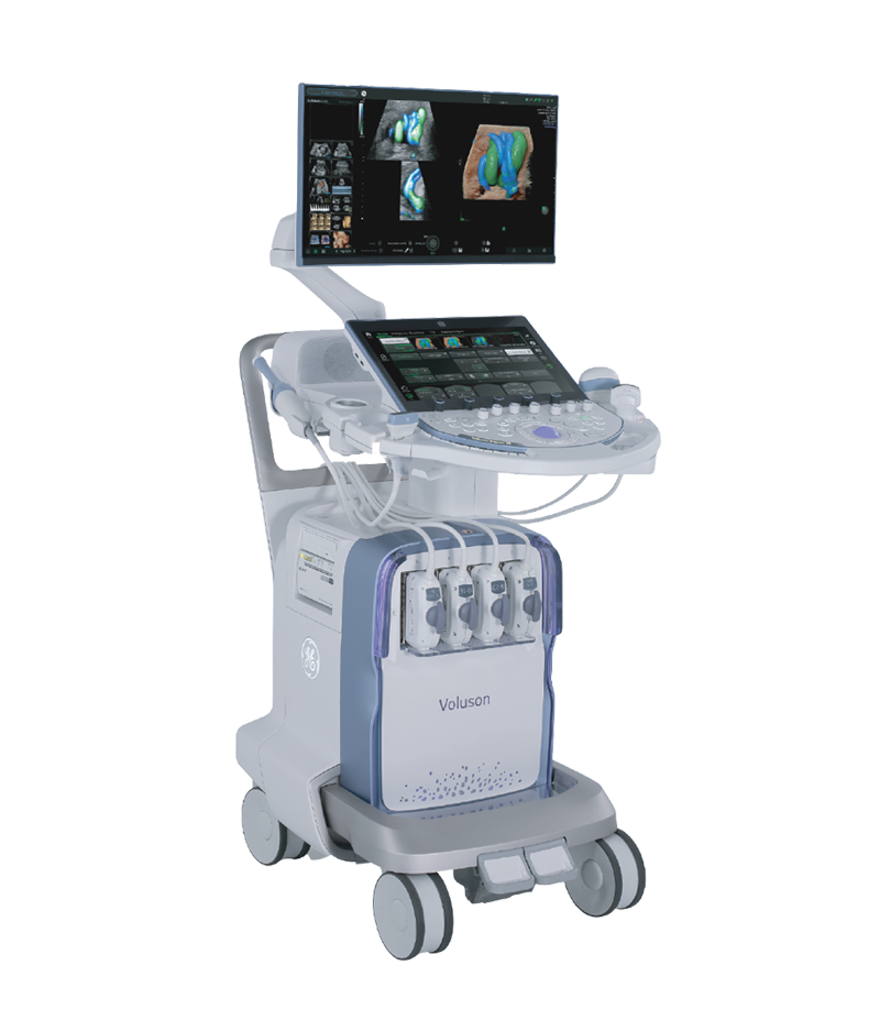




 Consult Now