 Chăm sóc khách hàng: 028 1234 5678
 Email: tdmedical@gmail.com


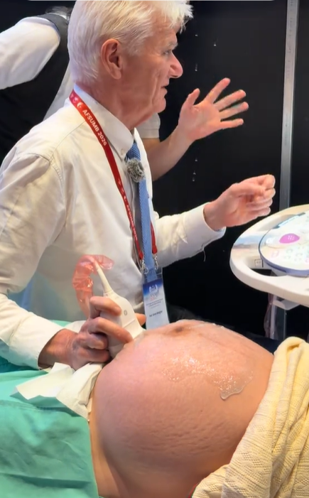
Professor Simon Meagher and Professor Ritsuko Pooh Performing Ultrasound Examinations Using the Voluson E22 System
 5.4K
5.4K
 17
17
 11-Sep-2026
 76,950
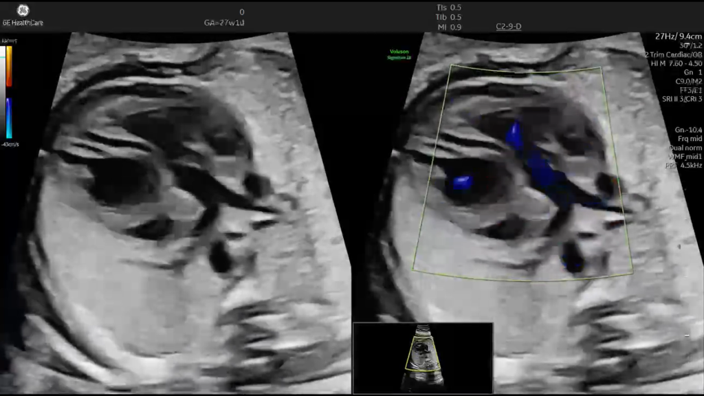
A 27-week-old fetus was referred for further evaluation after an abnormal orientation of the great arteries was detected during a routine ultrasound examination.
What initially appeared to be only a subtle anatomical variation quickly revealed much more intriguing and complex features when a detailed fetal echocardiographic examination was performed.
Let’s take a closer look at:
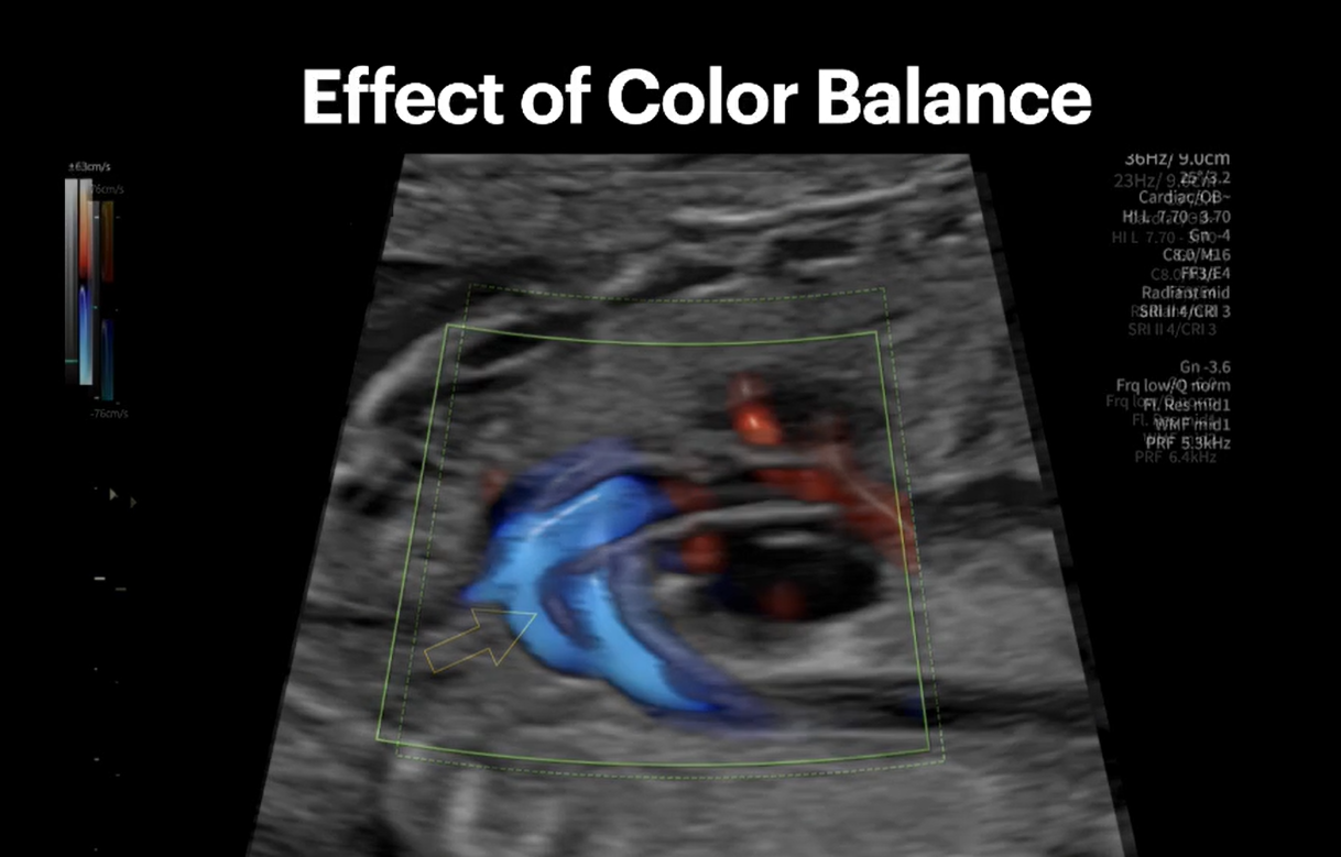
Something did not align with what we would normally expect to see in a structurally normal heart. The final diagnosis was a rare and severe congenital heart defect: congenitally corrected transposition of the great arteries (ccTGA). In this condition, the initial physiology may appear deceptively “balanced,” while the long-term clinical course can evolve very differently.
Video source: Dr. Shreepal
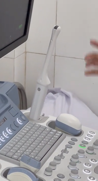
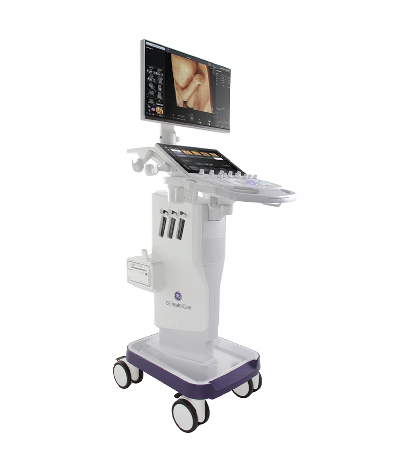
The ultrasound images were acquired using the Voluson Signature 18 system. Fetal anomalies, particularly congenital heart defects, are relatively uncommon but can have significant clinical consequences. Early and accurate detection allows physicians to develop more appropriate plans for further evaluation, management, and follow-up.
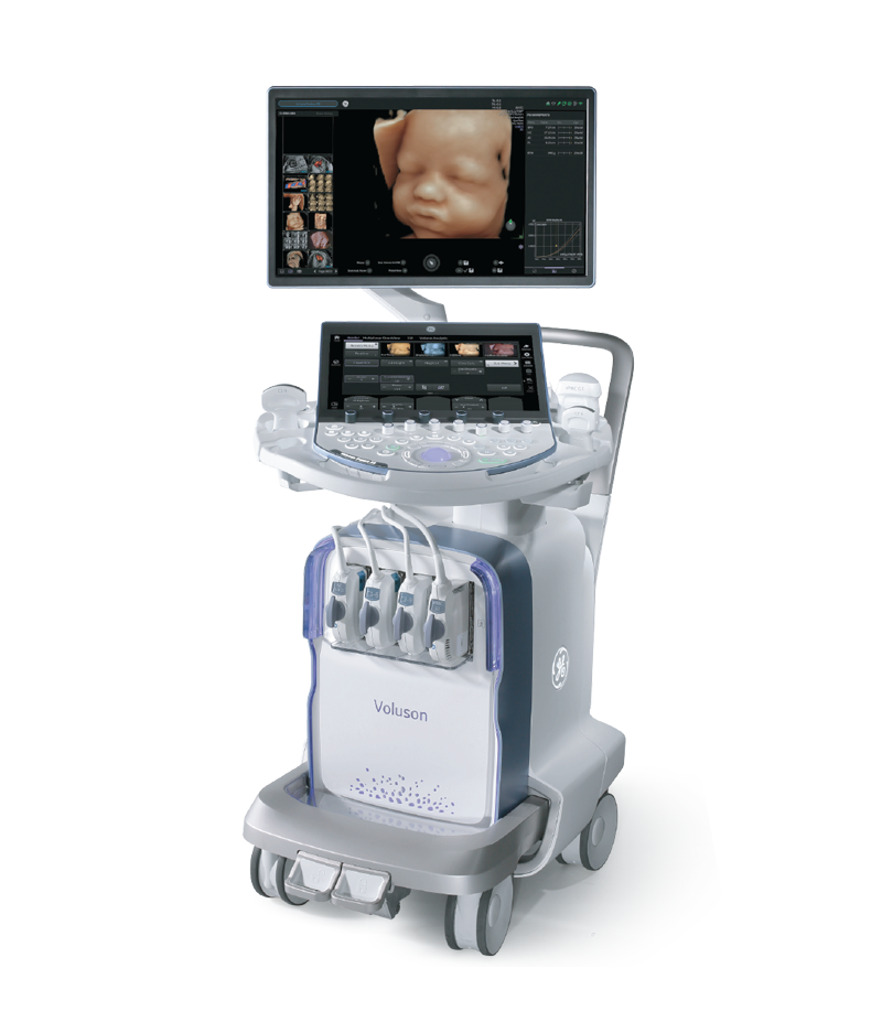
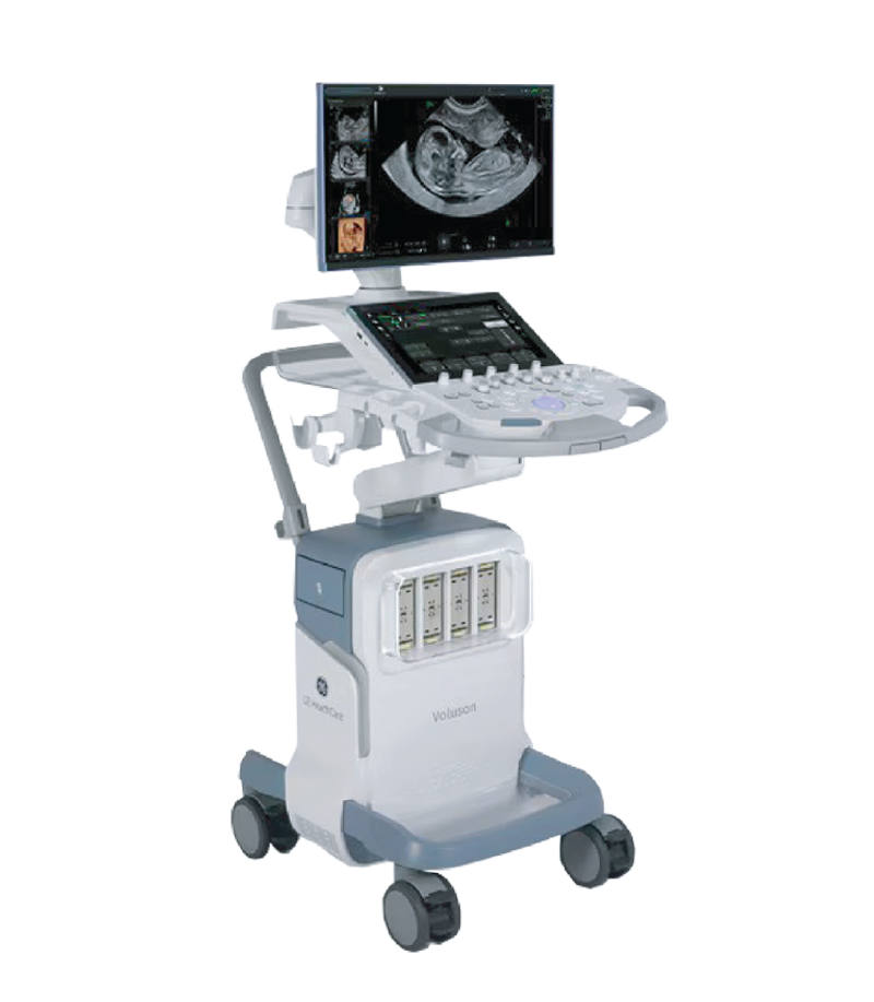
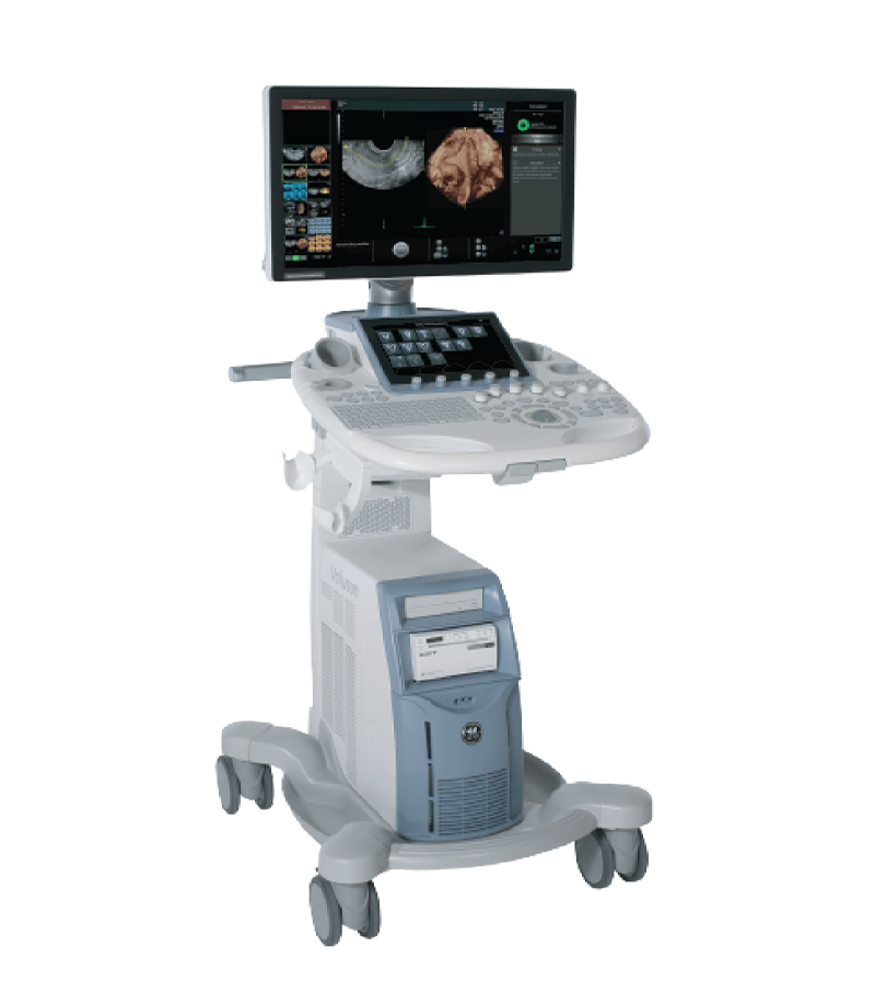
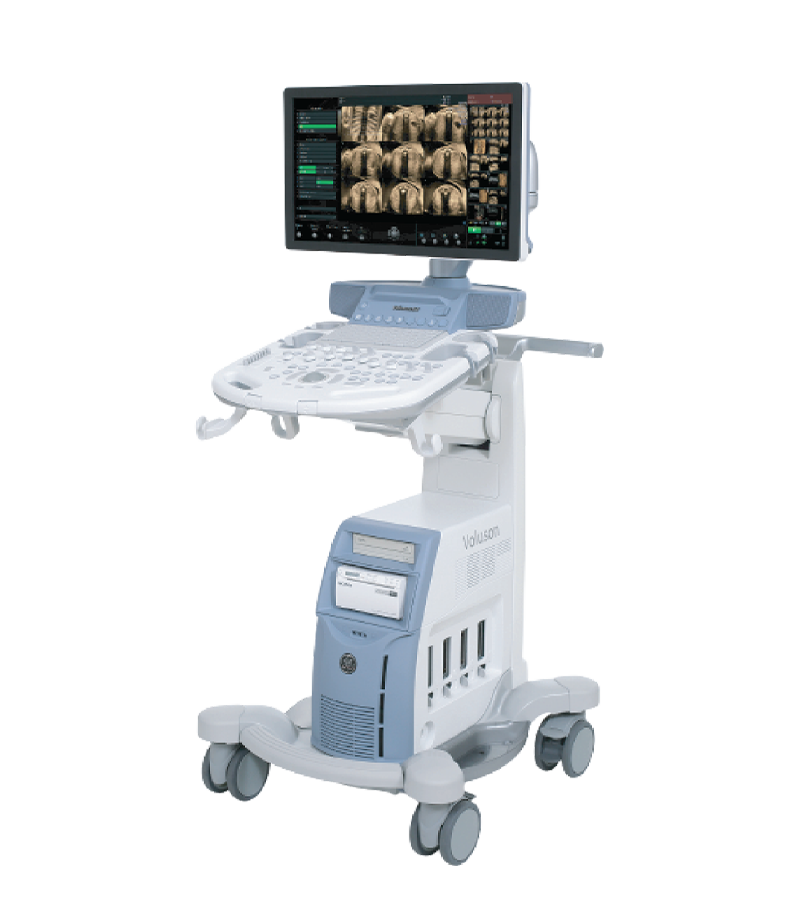
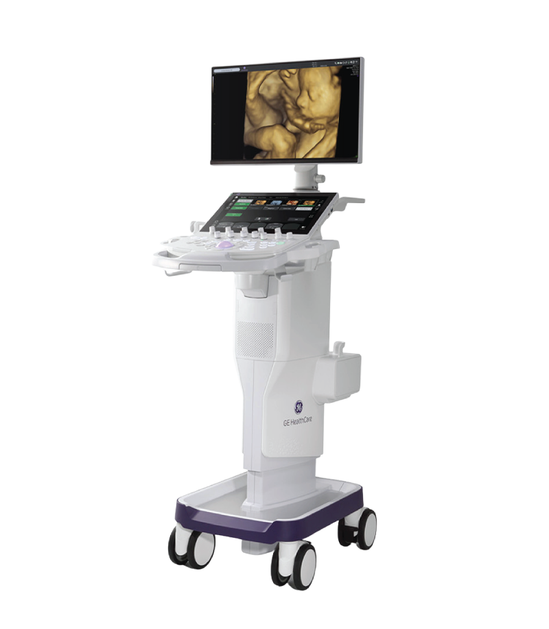
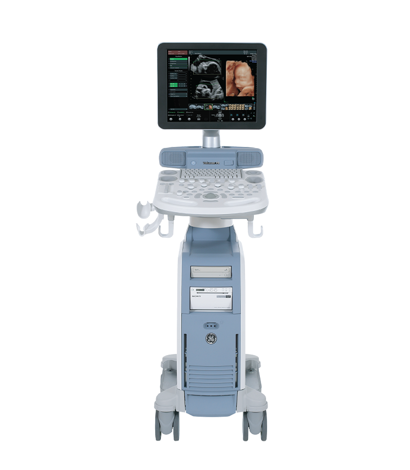
Voluson Ultrasound System
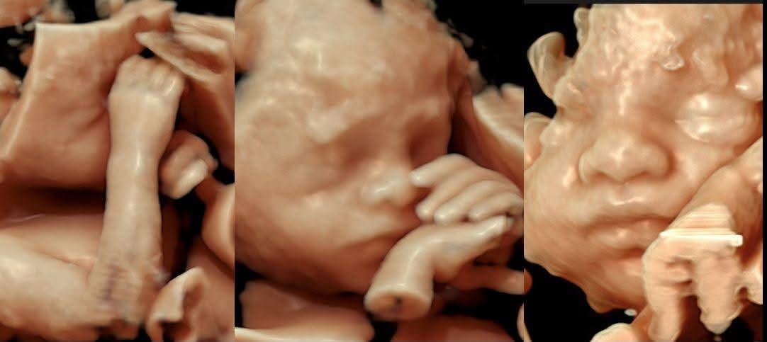
Voluson continues to strive to provide high-quality imaging and advanced technologies to support fetal screening and the detection of congenital abnormalities, helping minimize the risk of missed findings as much as possible.
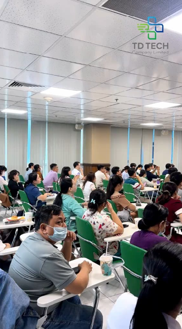
On March 20, 2026, TD Tech was honored to participate as a sponsor of the seminar “Energy and Nuclear Power in Vietnam,” held at the University of Science, Vietnam National University Ho Chi Minh City...
Xem thêm
The 9-month pregnancy journey is filled with a mix of emotions, from surprise and happiness to concerns about the baby’s development. If the first trimester is a time of adjustment and the second trimester brings...
Xem thêm
Congenital heart disease is among the most common congenital abnormalities in fetuses; however, prenatal detection remains challenging in routine obstetric practice. Although professional organizations such as ISUOG have issued detailed guidelines on the fetal cardiac...
Xem thêm
Every ultrasound examination offers more than just a glimpse of a baby’s anatomy—it also provides an opportunity to observe the remarkable activities taking place in real time. By around 27–28 weeks of pregnancy, the fetus...
Xem thêm
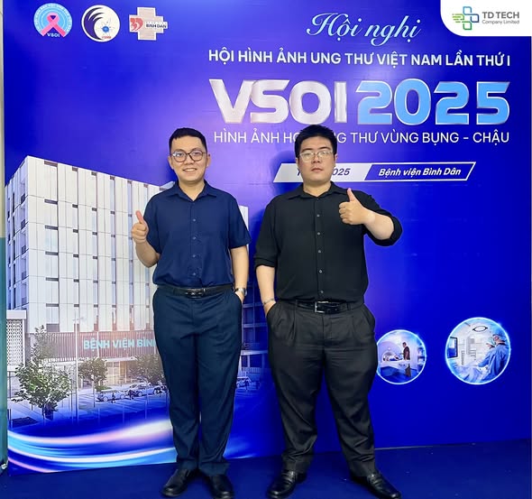
Comprehensive Medical Engineering Co., Ltd. was honored to accompany Binh Dan Hospital at the Vietnam Oncology Imaging Conference held on December 13. At the event, Toan Dien had the opportunity to introduce the RayStation Treatment...
Xem thêm
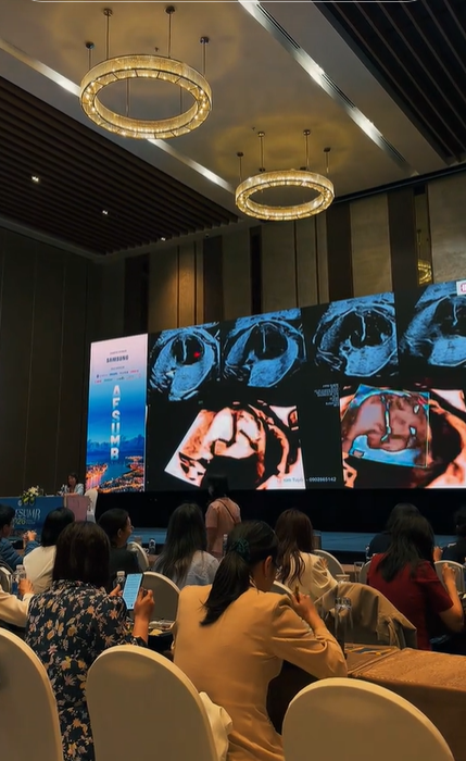
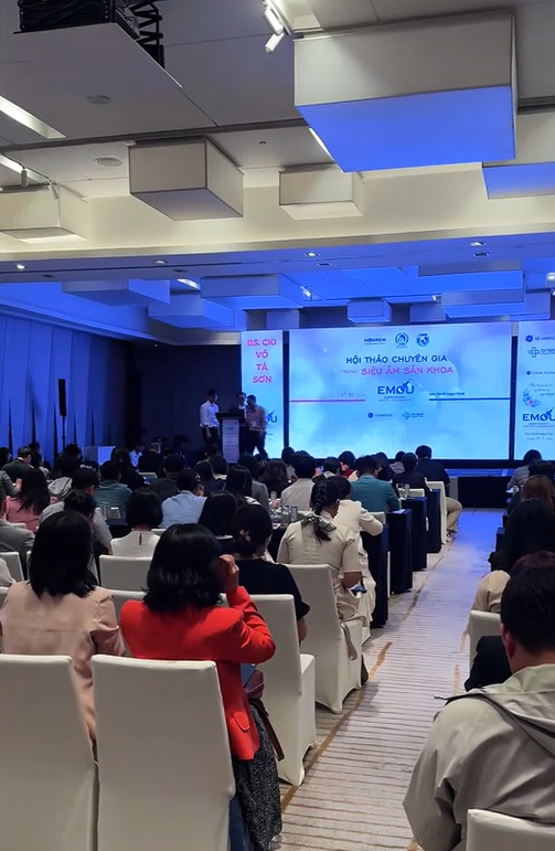
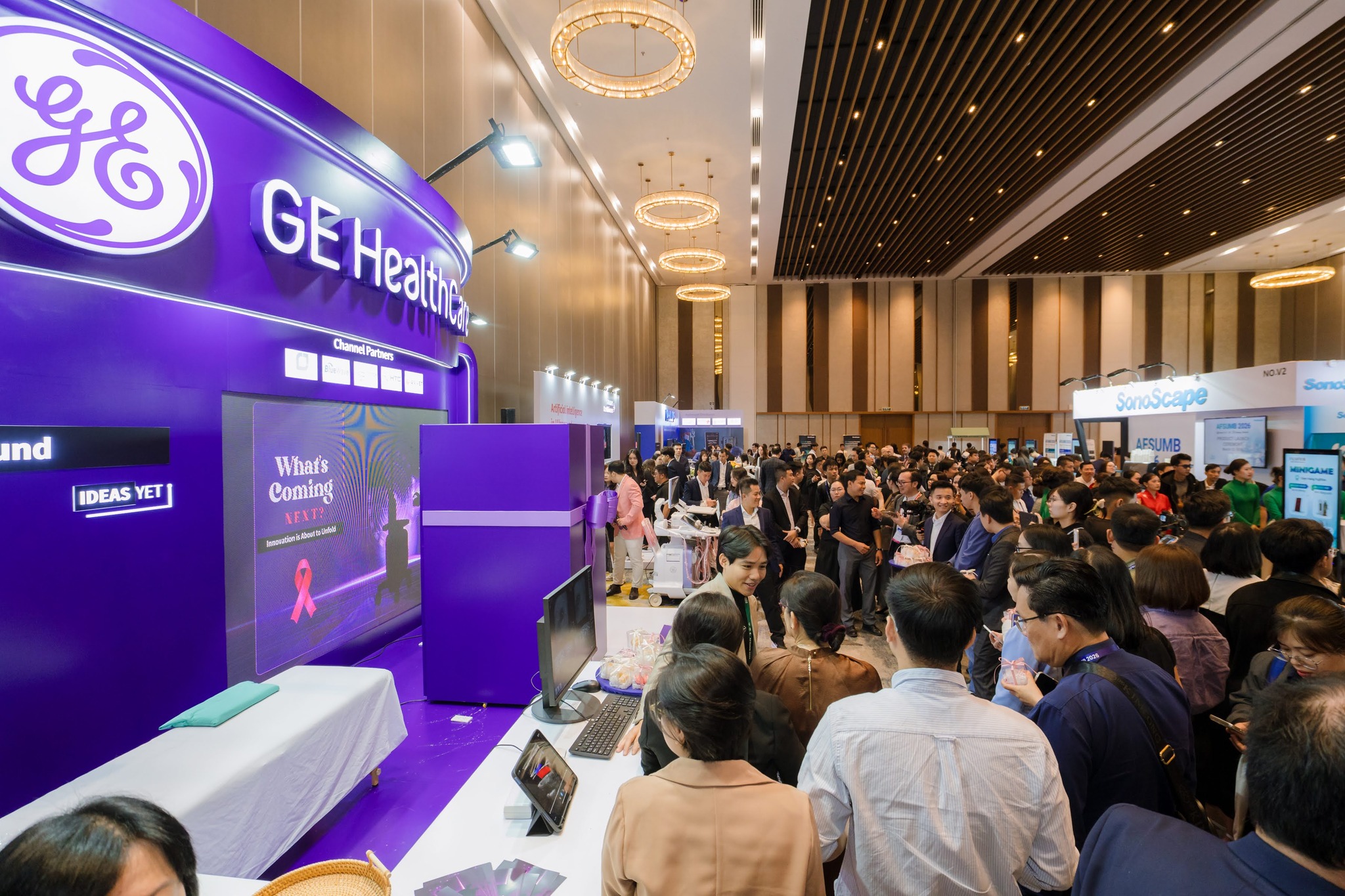
TD TECH was honored to partner with GE HealthCare as a Gold Sponsor at VSUM 2026—one of the largest scientific conferences in Vietnam, bringing together leading ultrasound experts, physicians, and sonographers from across the country....
Xem thêm

 Xem video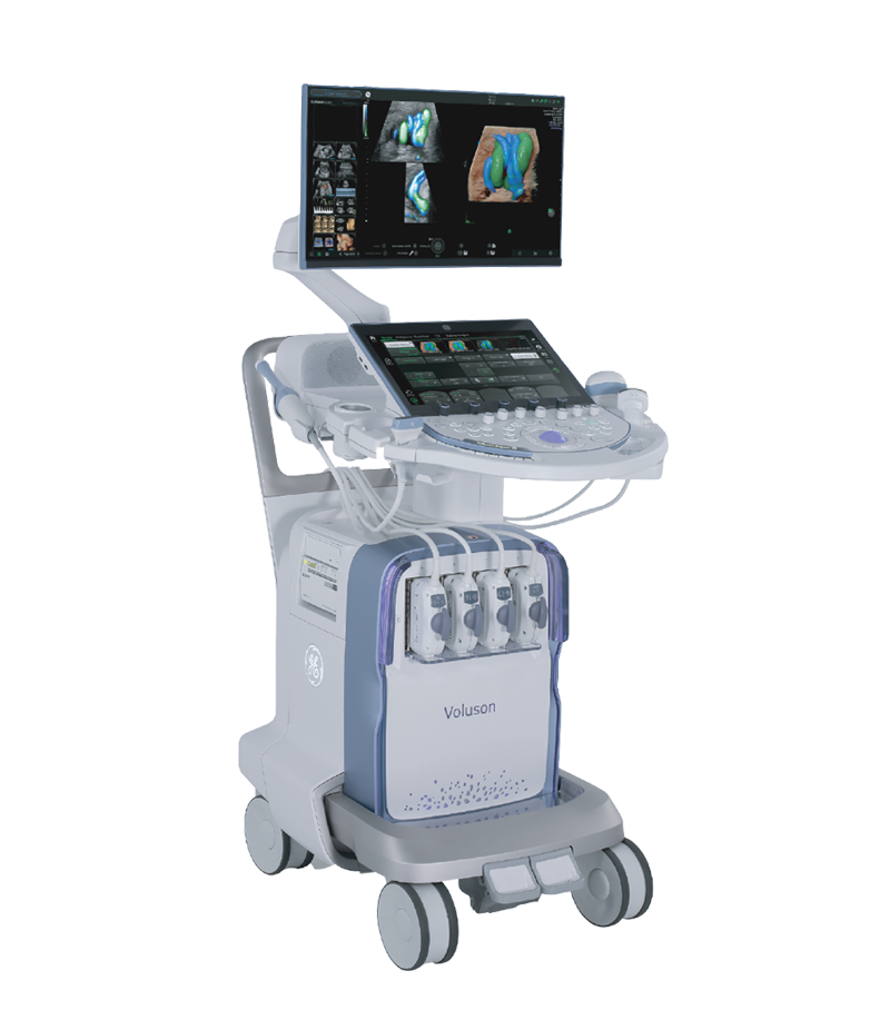




 Consult Now