 Email: tdmedical@gmail.com



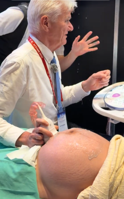
Professor Simon Meagher and Professor Ritsuko Pooh Performing Ultrasound Examinations Using the Voluson E22 System
 8.3K
8.3K
 51
51
 27-Aug-2026
 88,635
Congenital heart disease is among the most common congenital abnormalities in fetuses; however, prenatal detection remains challenging in routine obstetric practice. Although professional organizations such as ISUOG have issued detailed guidelines on the fetal cardiac ultrasound views that should be assessed, missed diagnoses still occur, mainly due to suboptimal image quality and inconsistent application of fundamental technical principles.
In a recent study published in Ultrasound in Obstetrics & Gynecology, the journal of ISUOG, Dr. Perez and colleagues introduced the DAZE technique (Depth, Angle, Zoom, Enhancement) – a systematic, easy-to-apply, and reproducible approach designed to optimize 2D ultrasound imaging as well as color Doppler in fetal cardiac assessment.
DAZE does not replace guidelines for fetal cardiac anatomical views. Instead, it serves as a standardized process to help the operator optimize image quality before assessing cardiac structure and function.
The fetal heart is a small structure that moves continuously at a high frequency and has complex anatomy. Image quality depends not only on the experience of the ultrasound operator but also closely on the adjustment of technical parameters on the ultrasound system.
Suboptimal imaging may reduce spatial resolution and temporal resolution, making it more difficult to identify small anatomical structures or abnormal blood flow on color Doppler. This directly affects the ability to detect fetal cardiac abnormalities, particularly complex congenital heart defects.
The DAZE technique was developed to standardize the image optimization process, enabling ultrasound operators to approach the examination in a more logical, sequential, and effective manner.
The first step of the DAZE technique is to optimize Depth and the focal zone. An excessively large depth setting causes the examination field to include unnecessary tissue, which can reduce frame rate and affect temporal resolution. Conversely, an excessively shallow depth may prevent the entire cardiac structure from being adequately visualized.
According to the DAZE principles:
This is the foundational step for improving image quality from the beginning of the examination.
“Angle” in the DAZE technique includes two important factors:
Reducing the scan angle width helps focus ultrasound energy on the region of interest, thereby increasing frame rate and improving temporal resolution. This is particularly important when assessing small structures such as heart valves, the interventricular septum, great vessels, and blood flow on color Doppler.
The direction of the ultrasound beam has a significant impact on image quality and the assessment of blood flow. In color Doppler, an appropriate examination angle helps display blood flow more clearly, reduce artifacts, and improve sensitivity for detecting hemodynamic abnormalities.
“Zoom” is the step that enhances visualization of small anatomical details. However, magnification is not simply a matter of making the image larger. The DAZE approach emphasizes the appropriate use of zoom when assessing standard fetal cardiac views, allowing clearer visualization of the heart valves, cardiac septa, and great vessels.
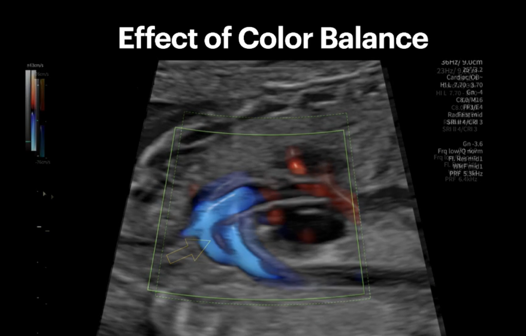
The final step of the DAZE technique is Enhancement – optimizing the settings of the ultrasound system. Parameters that may require adjustment include Gain, Dynamic range, Persistence, Speckle reduction, Harmonic imaging, Color Doppler settings, Pulse Repetition Frequency (PRF), and Wall filter.
For color Doppler, optimizing PRF, gain, and the color box is particularly important when evaluating blood flow across the heart valves, through the great vessels, and identifying abnormal flow patterns.
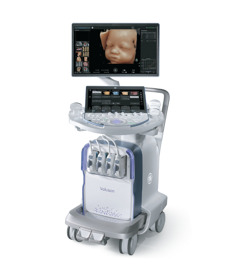
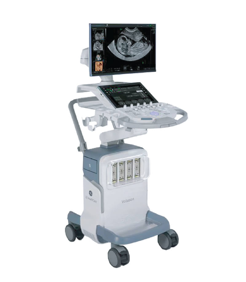
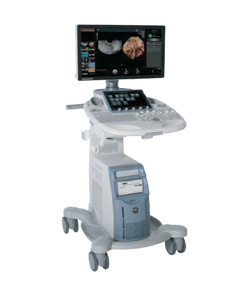
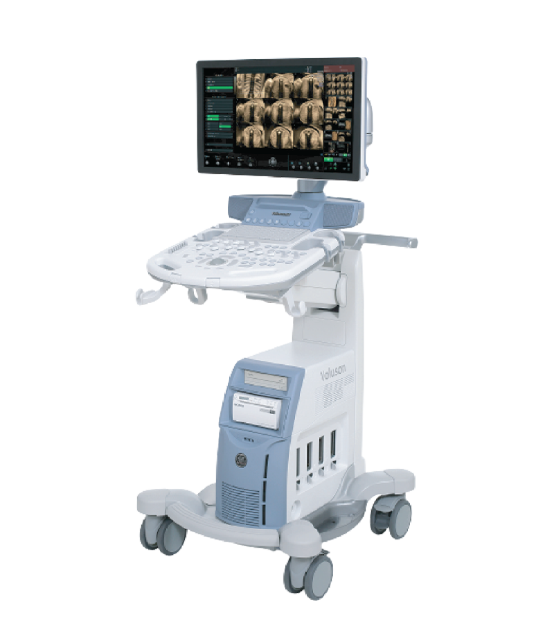
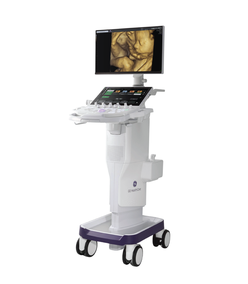
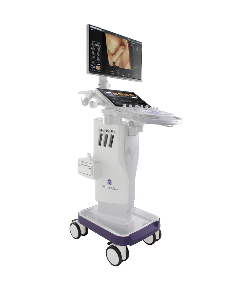
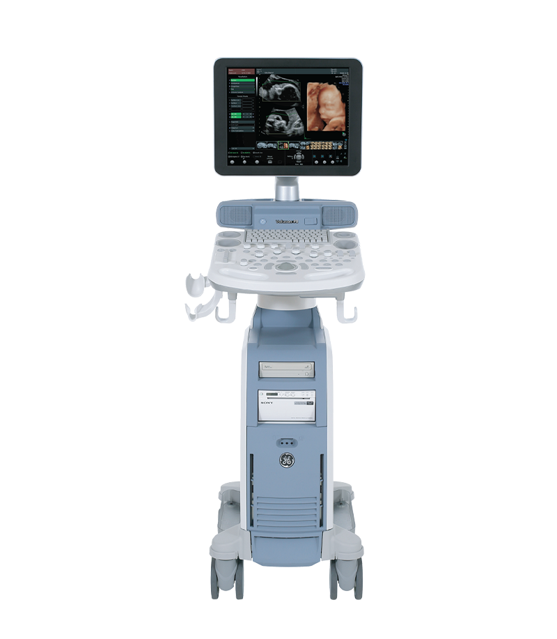
The study by Dr. Perez and colleagues used illustrative images and videos acquired using the Voluson ultrasound system from GE HealthCare.
Voluson Ultrasound System
With the ability to provide high-resolution imaging, optimize color Doppler, and support a range of advanced processing tools, modern ultrasound platforms play an important role in improving the quality of fetal cardiac assessment, particularly in the detection of congenital abnormalities.
As the need for prenatal diagnosis continues to grow, combining anatomical knowledge, practical scanning skills, and structured approaches such as DAZE may help improve screening effectiveness, support more accurate diagnosis, and provide practical benefits for both pregnant women and their fetuses.
Reference: https://obgyn.onlinelibrary.wiley.com/doi/10.1002/uog.70282
The same valve, the same ultrasound screen—yet they can tell three completely different stories. Video source: Dr. Shubham This video illustrates the appearance of the mitral valve in three different conditions: a normal mitral...
Xem thêm
With the latest cSound platform integrated, the Vivid™ cardiovascular ultrasound systems are delivering significant value in both clinical support and diagnostic imaging. This platform represents a major step forward in image quality, workflow smoothness, and day-to-day...
Xem thêm
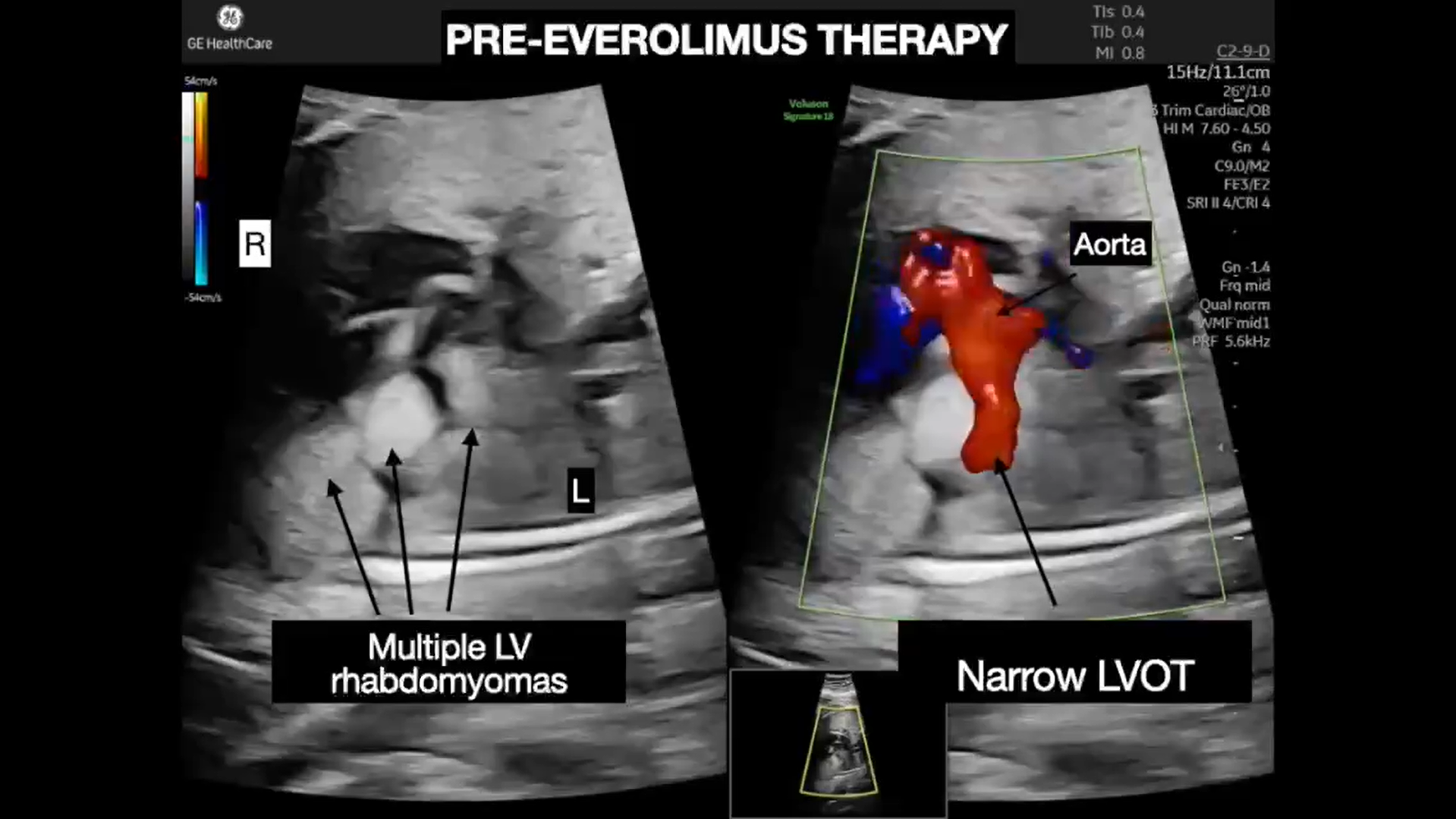
At 24 weeks of pregnancy, a routine screening ultrasound revealed multiple masses within the fetal heart. A subsequent fetal echocardiogram confirmed the presence of rhabdomyomas involving both ventricles. Although cardiac rhabdomyomas are generally benign...
Xem thêm
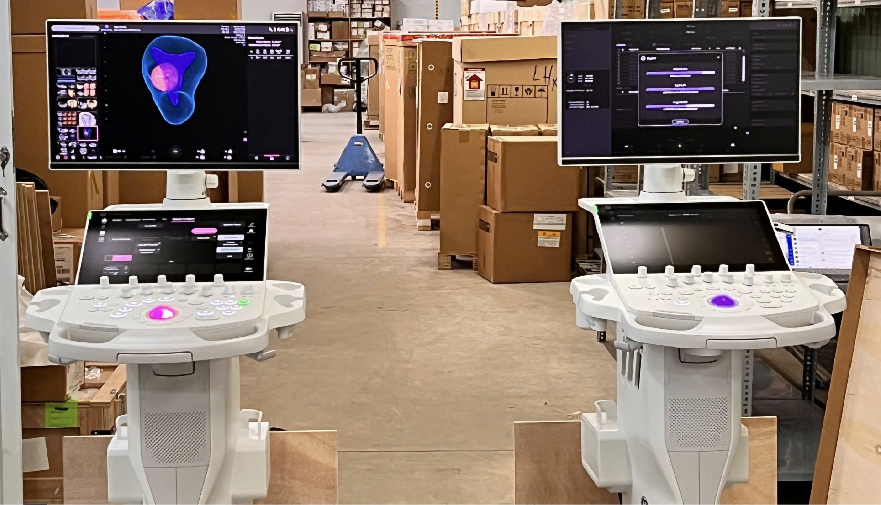
Unboxing the Voluson P16 and P18 ultrasound machines – the two latest models in the P-Series featuring modern designs, sharp imagery, and effective support for OB/GYN and multi-specialty diagnostics. Introduction In the field of diagnostic...
Xem thêm
During a transthoracic echocardiography (TTE) examination performed on a 21-year-old patient using a Vivid cardiovascular ultrasound system from GE HealthCare, an extracardiac mass in contact with the pericardium was identified. Further investigations subsequently led to...
Xem thêm
The 9-month pregnancy journey is filled with a mix of emotions, from surprise and happiness to concerns about the baby’s development. If the first trimester is a time of adjustment and the second trimester brings...
Xem thêm





 Xem video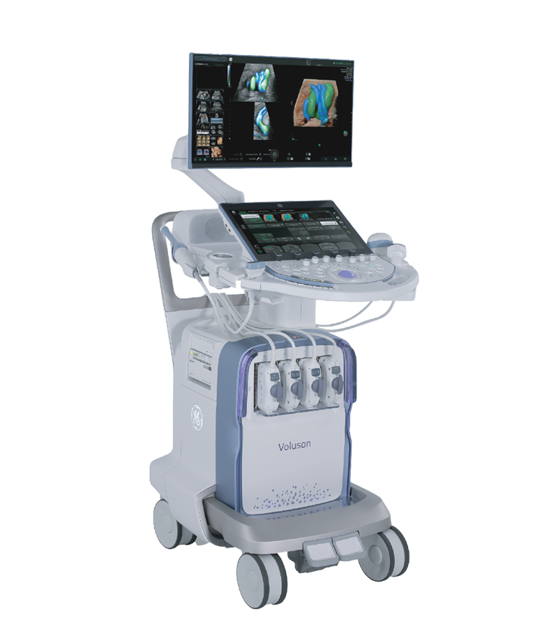



 Consult Now