

1. Imaging Technology – The Solid Foundation of Voluson P8
Despite being in the economic segment, Voluson P8 is still equipped by GE with many important imaging technologies:
✔ Sharp 2D Imaging – High Contrast
-
Excellent soft tissue display
-
Clear anatomical boundaries
-
Reduced noise
-
Easy observation of early pregnancy and gynecological structures
✔ Flexible 3D/4D Ultrasound
While not a premium 4D line, Voluson P8 still supports 4D rendering for:
-
Fetal face rendering
-
Observation of limbs and spine
-
3D uterine structure display
Smooth, natural imaging suitable for patient consultation and recording.
✔ Speckle Reduction Imaging (SRI) – Image Noise Reduction
SRI helps to:
-
Eliminate artifacts
-
Smooth images while preserving tissue details
-
Optimize for difficult-to-scan patients
✔ CrossXBeam™ – Contrast Enhancement
Combines multiple scan angles to:
-
Highlight boundaries
-
Increase soft tissue sharpness
-
Provide strong support in gynecological and pregnancy surveys
✔ Sensitive Color and Spectral Doppler
Voluson P8 supports:
-
Color Doppler
-
Power Doppler
-
Spectral Doppler
Used for:
-
Fetal heart surveys
-
Placental blood flow
-
Uterine flow
-
Ovarian or cyst perfusion
2. Clinical Applications – Comprehensive for Obstetrics & Gynecology
2.1 Early Pregnancy – Clear Imaging from the First Weeks
Voluson P8 helps clinicians:
-
Observe the gestational sac
-
Check intrauterine pregnancy position
-
Evaluate early embryos
-
Monitor fetal heart during the first 6–8 weeks
Bright images with low noise help in early detection of abnormalities.
2.2 Pregnancy Survey – Daily 2D & Flexible 4D
P8’s pregnancy survey capabilities are ideal for:
-
Routine pregnancy checkups
-
Fetal morphology assessment
-
4D fetal face observation
-
Observation of limbs, spine, and internal organs
Smooth 4D imaging well meets consultation and fetal monitoring needs.
2.3 Fetal Heart – Effective Assessment for Clinics
Voluson P8 supports basic fetal heart assessment:
-
Clear Doppler
-
Standard heart views
-
Evaluation of valves and chambers
-
Real-time heart rate observation
Suitable for OB/GYN clinics and district-level hospitals.
2.4 Gynecology – Comprehensive Survey of Uterus & Ovaries
P8 helps clinicians detect:
-
Uterine fibroids
-
Ovarian cysts
-
Endometrial polyps
-
Uterine malformations
-
IVF endometrial assessment
3D imaging technology helps clearly reconstruct the uterus, useful in infertility diagnosis and pre-surgery.
2.5 IVF Support – Cost-effective Solution for Infertility Clinics
Voluson P8 effectively meets basic IVF needs:
-
Antral Follicle Count (AFC)
-
Stimulation cycle monitoring
-
Endometrial evaluation
-
Ultrasound-guided intervention support
Helps IVF centers save on initial investment while ensuring efficiency.
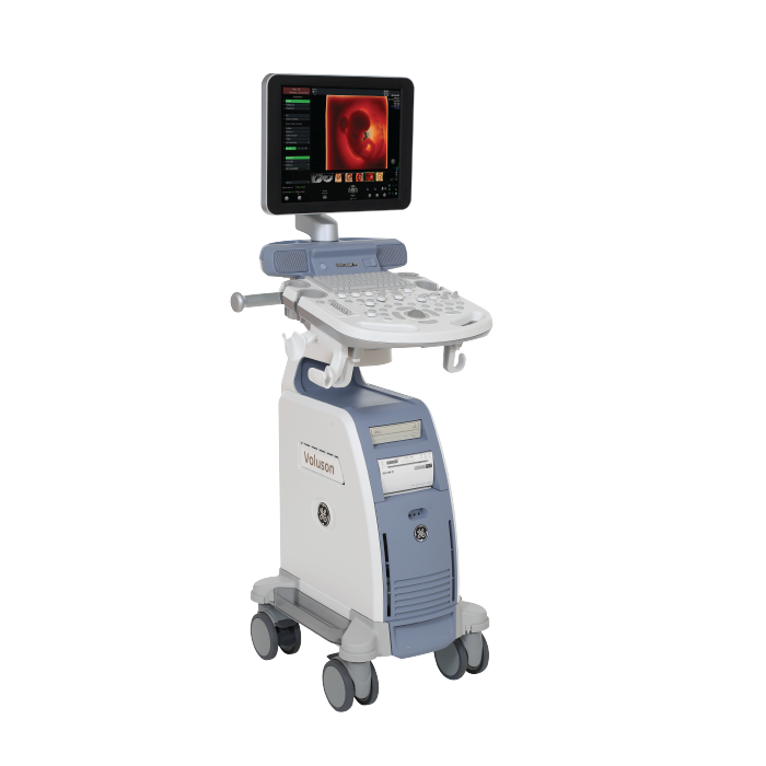
3. User-friendly Design – Easy to Use for Every Clinician
A major strength of the Voluson P8 is its simple design and ease of operation:
✔ Intuitive Interface
Functional keys are logically arranged, easy to learn and operate.
✔ Clear and Bright Display
Better imaging even in high-light environments.
✔ Diverse Probes
Supports abdominal, vaginal, thyroid, breast, and soft tissue probes...
✔ Compact – Flexible
Easy to move within the clinic or from room to room.
✔ Modern Connectivity
Compatible with PACS – DICOM – USB – high-quality image/video export.
4. Investment Benefits – Economical yet Durable Choice
Voluson P8 is an equipment aimed at:
-
Lowest price in the Voluson P-Series line
-
Easy maintenance – High durability
-
Low operating costs
-
Fast Return on Investment (ROI)
-
Increased diagnostic productivity
P8 is highly suitable for:
-
Newly opened private clinics
-
District/Provincial hospitals
-
Women's health centers
-
Mobile ultrasound units

