

1. Advanced Imaging Technology – The Foundation of Voluson S8t’s Superiority
✔ High-Resolution 2D
Sharp 2D imaging and strong contrast allow clinicians to easily observe soft tissue, the fetus, and small structures such as the endometrium and ovarian follicles.
✔ HDlive™ – Realistic 4D Imaging
HDlive technology simulates natural light and shadows, providing lifelike and vivid 4D images for:
-
Fetal face observation
-
Surveying limbs, spine, and organs
-
Enhancing visual intuition during patient counseling
✔ SRI (Speckle Reduction Imaging) – Noise reduction while preserving detail
Particularly useful when:
-
Scanning high-BMI patients
-
Surveying soft tissue or difficult-to-visualize areas
✔ CrossXBeam™ – Contrast Enhancement
Supports clear visualization of tissue boundaries, aiding in the assessment of cysts, the endometrium, ovaries, and fetal structures.
✔ Powerful Doppler
Includes Color, Power, and Spectral Doppler to assist in surveying:
-
Fetal heart
-
Placental vasculature
-
Uterine arteries
-
Ovaries & soft tissue perfusion
2. Clinical Applications – Versatile and Precise for OB/GYN
2.1 Early Pregnancy & Pregnancy – Comprehensive Diagnostic Support
Voluson S8t supports:
-
Embryo survey from weeks 6–8
-
NT (Nuchal Translucency) measurement
-
Morphology assessment at all stages
-
Detailed observation of face, limbs, brain, and fetal heart
-
Monitoring pregnancy development
-
Smooth, high-quality 4D rendering
Bright, clear, and low-noise imaging helps clinicians detect early signs of abnormalities.
2.2 Fetal Heart – Sensitive Doppler, Sharp Imaging
Voluson S8t is a suitable choice for fetal heart surveys thanks to:
-
High-sensitivity Doppler
-
Display of standard heart views (4-chamber, 3-vessel, aortic arch)
-
Real-time heart rate observation
-
Evaluation of abnormal flows
This assists in the early detection of common congenital heart diseases.
2.3 Gynecology – Clear Visualization of the Uterus and Ovaries
The system supports effective diagnosis of:
-
Uterine fibroids
-
Endometrial polyps
-
Ovarian cysts
-
PCOS (Polycystic Ovary Syndrome)
-
Uterine anomalies (septum, bicornuate, etc.)
-
Endometrial assessment for IVF
VCI technology and 3D/4D imaging help reconstruct tumors and soft tissue structures more clearly.
2.4 IVF Support – A Crucial Tool for Infertility Clinics
Voluson S8t supports:
-
Antral Follicle Count (AFC)
-
Follicle monitoring
-
Endometrial evaluation
-
Precise procedural guidance
IVF clinicians value the S8t for its stability and sensitivity to small structures.
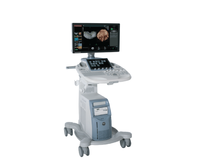
3. Design – Optimized for Busy Clinics
Voluson S8t is designed to help clinicians operate quickly and accurately:
✔ Intuitive Interface, Easy to Use
Reduces learning curves and operation time.
✔ Sharp Display Monitor
Allows for clear observation of small tissue details and Doppler signals.
✔ Diverse Probes
Supports abdominal, vaginal, thyroid, cardiac probes, and more.
✔ Fast Processing Speed
Helps increase the number of daily examinations.
✔ Modern Integrated Connectivity
DICOM – PACS – High-quality image/video export.
4. Investment Benefits – Superior Economic Value for Medical Facilities
Voluson S8t is an attractive choice because of:
-
More reasonable investment cost compared to the S10 Expert or E-Series
-
Powerful performance → shortening ultrasound time
-
Beautiful imaging → enhancing clinic reputation
-
Easy maintenance, high durability
-
Optimized for private clinics, district/provincial hospitals
Many facilities choose the S8t for its fast Return on Investment (ROI) and high satisfaction levels from both clinicians & patients.

