

1. Powerful Technological Foundation – Creating Superior Image Quality
Voluson P18 applies prominent GE technologies to optimize resolution and enhance detailed observation capabilities.
✔ Sharp 2D Imaging – High Contrast
-
Clear display of tissue structures
-
Sharp anatomical boundaries
-
Very useful for assessing early pregnancy, soft tissue, and gynecological structures
✔ HDlive™ – The Gold Standard of 4D Imaging
Voluson P18 supports HDlive, which helps to:
-
Generate natural and realistic fetal face imaging
-
Reproduce precise lighting and shadows
-
Support intuitive visual counseling for patients
✔ SRI Noise Reduction Technology
Speckle Reduction Imaging helps to:
-
Eliminate image artifacts
-
Smooth the image without losing detail
-
Optimize for high-BMI patients
✔ CrossXBeam™ – Enhancing Tissue Contrast
Allows clear display of tissue boundaries, supporting:
-
Fibroid assessment
-
Ovarian observation
-
More accurate endometrial measurement
✔ High-Sensitivity Doppler – Supporting Fetal Heart and Gynecology
Voluson P18 Doppler includes:
-
Color Doppler
-
Power Doppler
-
Spectral Doppler
Allows for the assessment of:
-
Fetal heart
-
Placental vasculature
-
Uterine arteries
-
Ovarian perfusion
2. Comprehensive Clinical Applications – Optimized for Obstetrics, Gynecology & IVF
2.1 Early Pregnancy – Detailed Survey from the First Weeks
Voluson P18 supports:
-
Observation of the embryo from weeks 6–7
-
Verification of gestational sac position
-
Assessment of early fetal heart activity
-
Accurate NT (Nuchal Translucency) measurement in first-trimester screening
Bright and clear imaging helps in early detection of pregnancy abnormalities.
2.2 Comprehensive Fetal Morphology Survey
The system provides the capability to assess:
-
Brain, spine, heart, lungs
-
Fetal face (nose, lips, jaw bone...)
-
Limbs and long bone structures
-
Internal organs using 4D
Smooth 4D imaging allows clinicians to observe comprehensively and in detail.
2.3 Fetal Heart – Reliable Assessment
Even without STIC like the Expert series, Voluson P18 remains very powerful in fetal heart imaging thanks to:
-
Sensitive Doppler with clear flow display
-
Standard 4-chamber and 3-vessel views
-
Sharp display of cardiac valve and chamber structures
-
Early detection of abnormal signs
2.4 Gynecology – Sharp Display of Uterus & Ovaries
Voluson P18 is ideal for gynecological surveys:
-
Endometrium
-
PCOS (Polycystic Ovary Syndrome)
-
Uterine fibroids
-
Ovarian cysts
-
Uterine polyps
-
Uterine malformations
3D/4D imaging helps precisely assess uterine shape and lesion locations.
2.5 IVF Support – Essential Tool for Infertility Clinics
Voluson P18 supports:
-
AFC (Antral Follicle Count) measurement
-
Monitoring follicles during stimulation cycles
-
Assessing endometrial thickness & structure
-
Supporting IVF procedures under ultrasound guidance
This is a popular choice for assisted reproductive centers.
3. User-Friendly Design – Optimized Workflow
Voluson P18 is designed to help clinicians operate faster and more accurately.
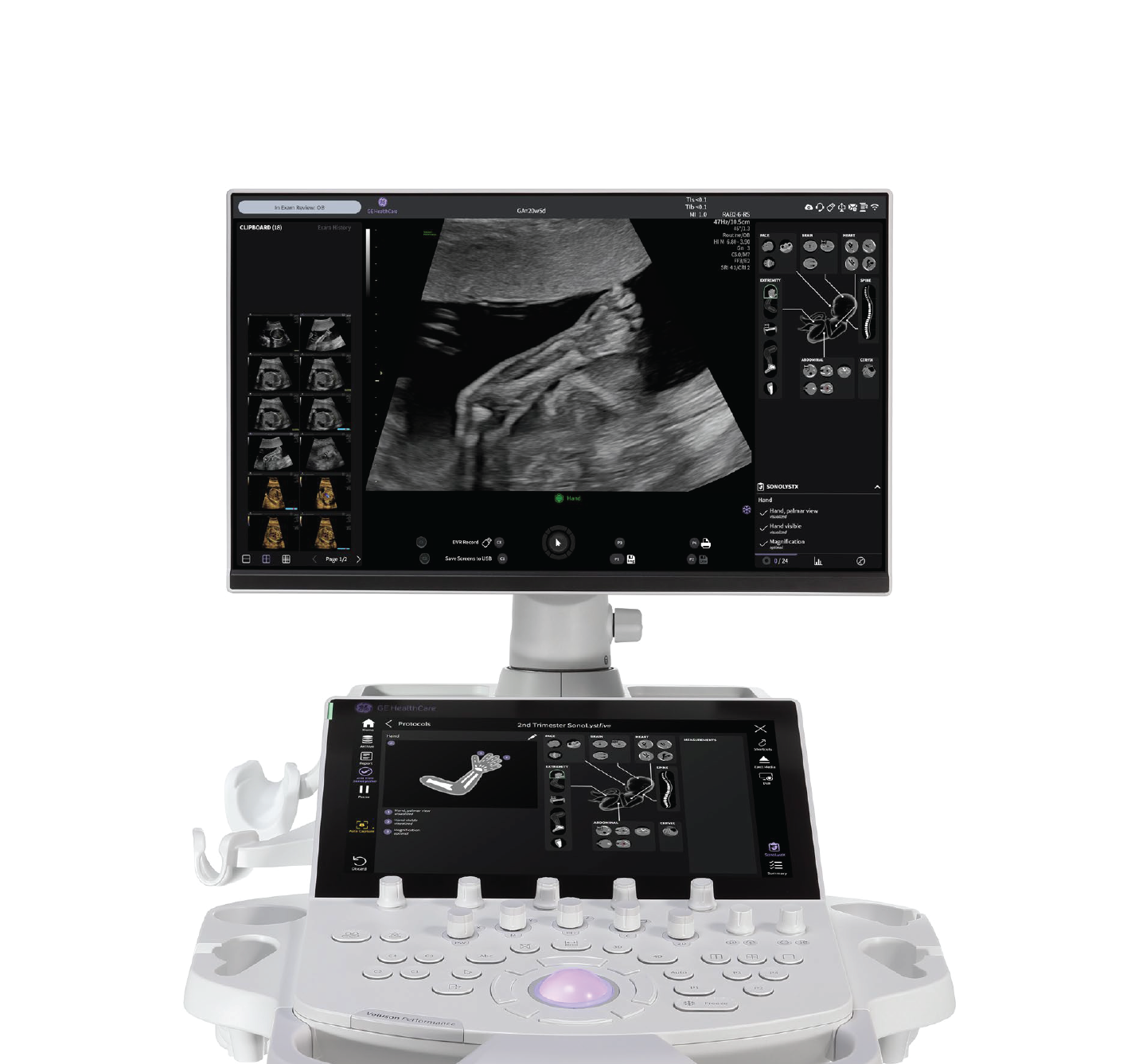
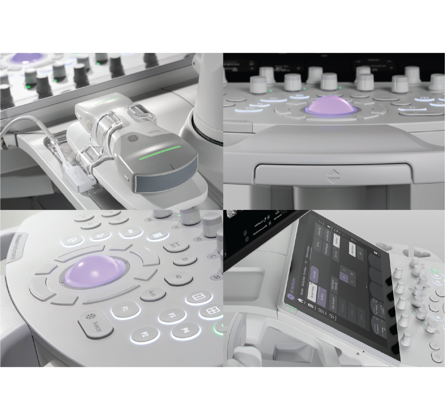
✔ Intuitive Interface
Logical control button layout — easy to learn, easy to use.
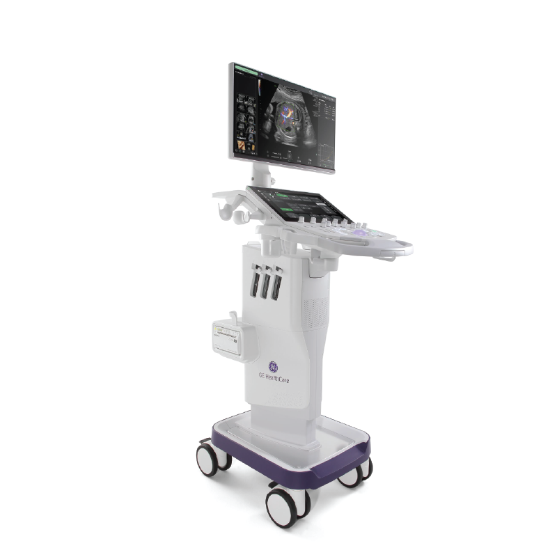
✔ Large, Sharp Display Monitor
Enables better observation of small anatomical structures.
✔ Smart Keyboard and Shortcuts
Supports rapid examinations and reduces redundant actions.
✔ Support for Multiple Probes
Suitable for:
-
Pregnancy scans
-
Gynecological exams
-
Thyroid, breast, and soft tissue exams
-
Fetal cardiology
✔ DICOM/PACS Compatibility
Easily connects with hospital information systems.
4. Economic Benefits – Smart Investment for Medical Facilities
Voluson P18 is a worthy investment thanks to:
-
Mid-to-high level cost but with specialized features
-
High durability and minimal downtime
-
Beautiful imaging -> increasing patient satisfaction
-
Fast ultrasound workflow -> increasing examination throughput
-
Excellent Return on Investment (ROI)
Suitable for:
-
Premium OB/GYN clinics
-
IVF facilities
-
District and provincial hospitals
-
Women's health centers

