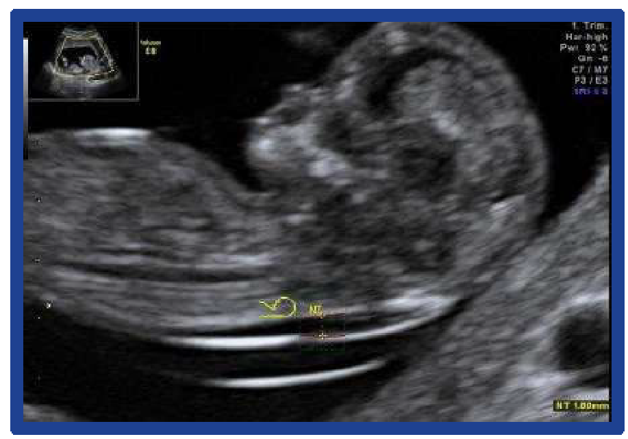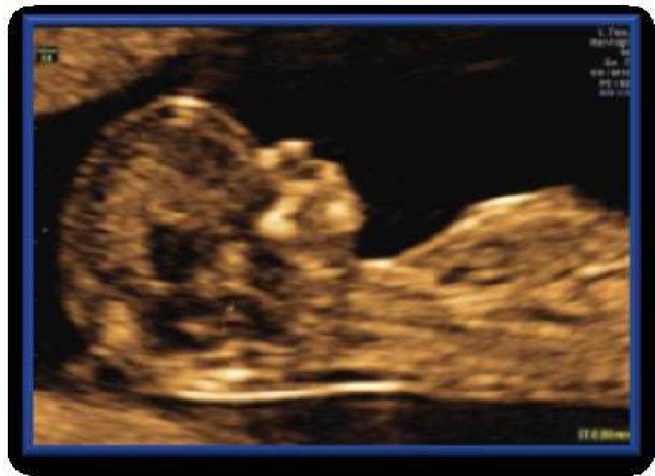
Behind every high-quality diagnostic image are advanced imaging technologies and artificial intelligence developed by GE HealthCare over generations of innovation. From HDlive for lifelike 3D/4D rendering to the Edison AI platform, TD Tech brings these cutting-edge technologies to hospitals and clinics across Vietnam.
Proprietary Imaging Technologies: The Soul of the Voluson Line
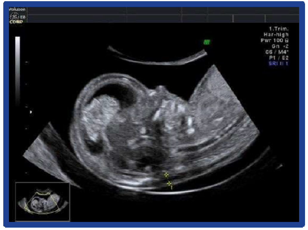
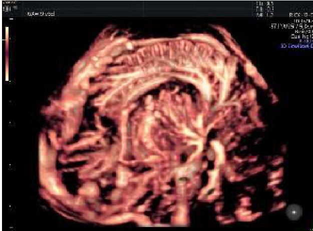
HDlive™: The 3D/4D Revolution
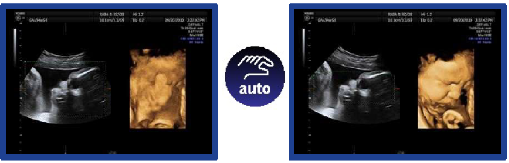
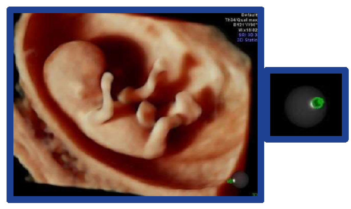
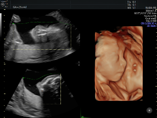
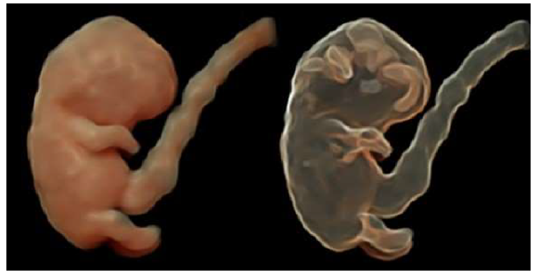
HDlive is not merely 3D rendering, but the use of virtual light sources and shadows to create superior depth and tissue detail.
HDlive Studio+: Combines three independent light sources to display the fetal face with lifelike realism.8
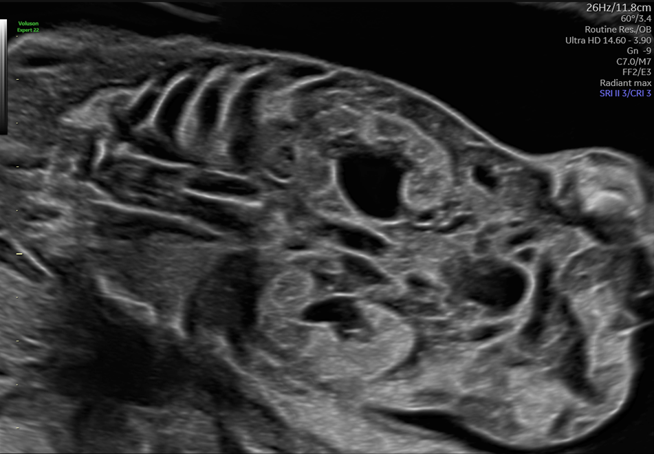
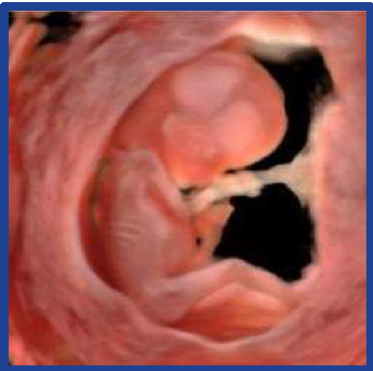
HDlive Silhouette: Helps physicians see through the outer tissue layers to observe the internal structure of the embryo or internal organs.
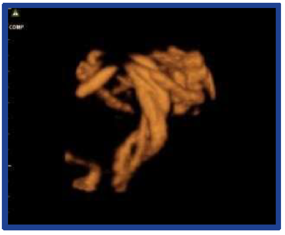
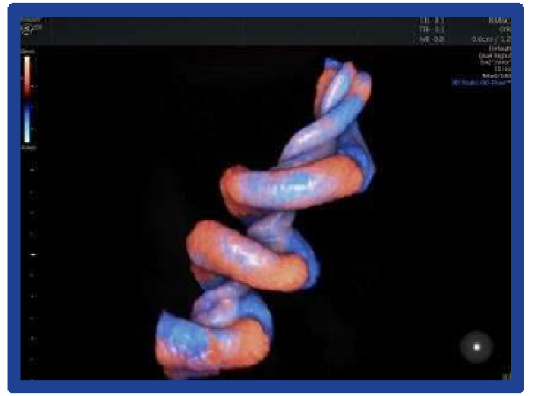
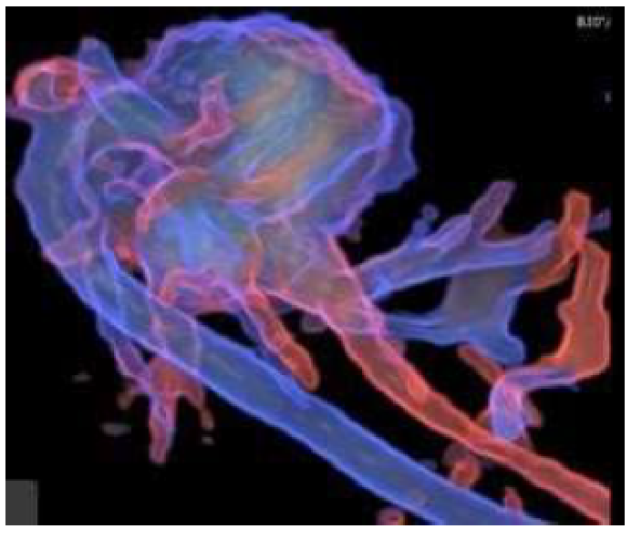
HDlive Flow: Integrates blood-flow data into the 3D volume-rendered image, helping clearly visualize the development of the fetal vascular system
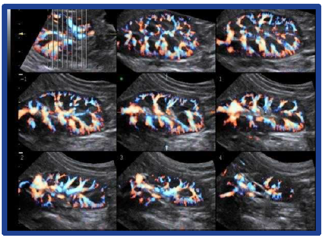
Radiantflow™ And SlowflowHD
GE HealthCare has redefined color Doppler ultrasound with these technologies:
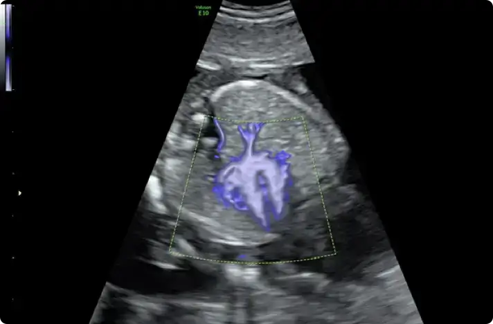
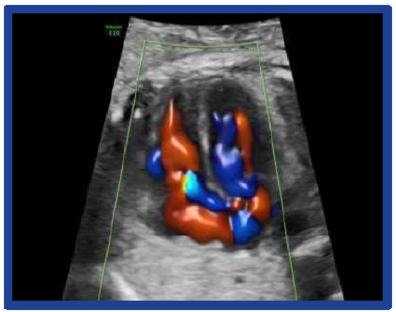
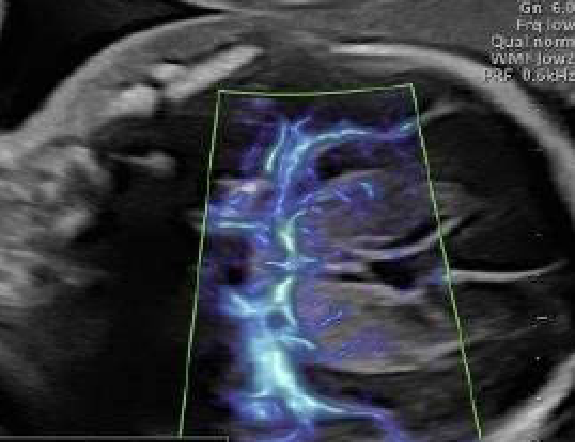
Radiantflow: Uses the amplitude of the color Doppler signal to create flow images with 3D depth. This makes it easy for physicians to identify tiny blood vessels and the boundaries between them, especially important in evaluating the fetal heart and cerebral vessels.
SlowflowHD: Extends visualization down to extremely slow flows, allowing assessment of perfusion within tumors or microcirculation in the ovaries and endometrium.
Augment And Shadow Reduction
These features are designed to solve the everyday challenges of the sonographer:
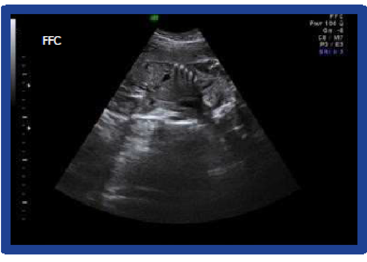
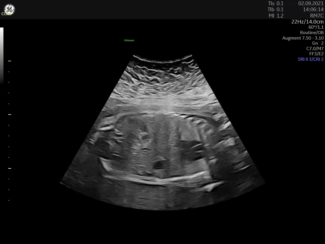
Augment: With just one button, the system automatically reduces noise and enhances penetration, significantly improving image quality in patients with a high BMI.
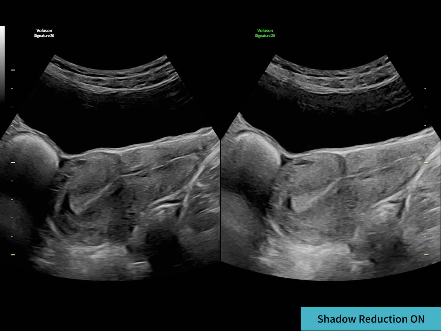
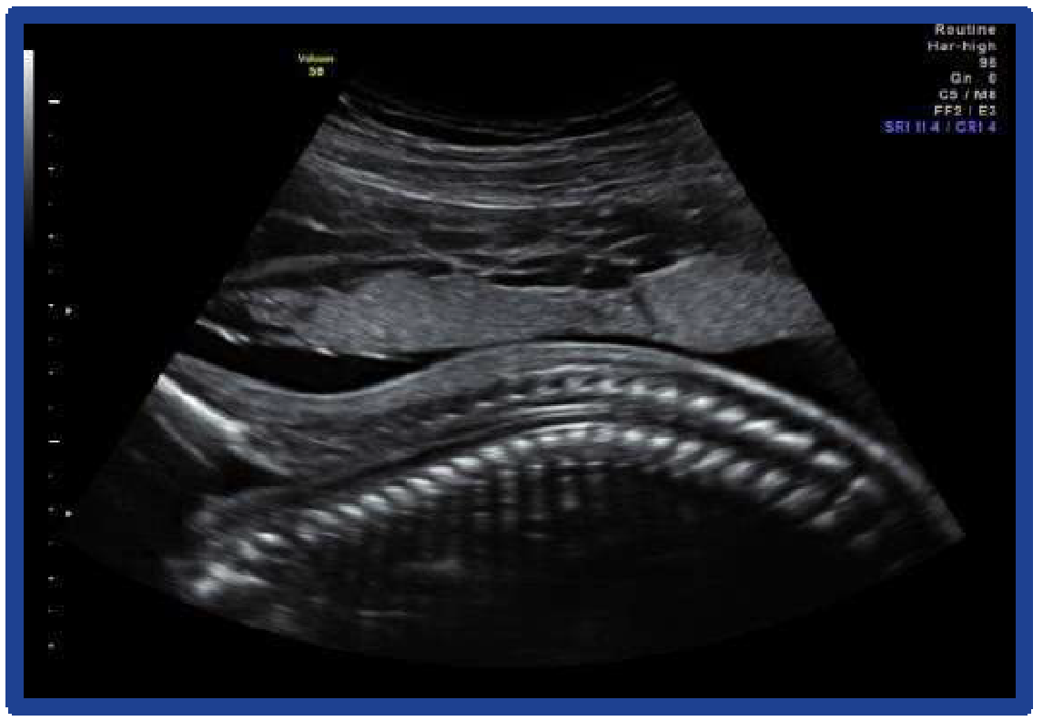
Shadow Reduction: Automatically compensates for shadowing behind bony structures (such as the ribs or fetal spine), helping restore obscured tissue information so physicians get a more complete view
Artificial Intelligence And Automation: The Edison AI Trend
GE HealthCare has deeply integrated AI into the Voluson lines through the Edison platform, helping standardize results and reduce operator fatigue.
SonoLyst: The Virtual Assistant
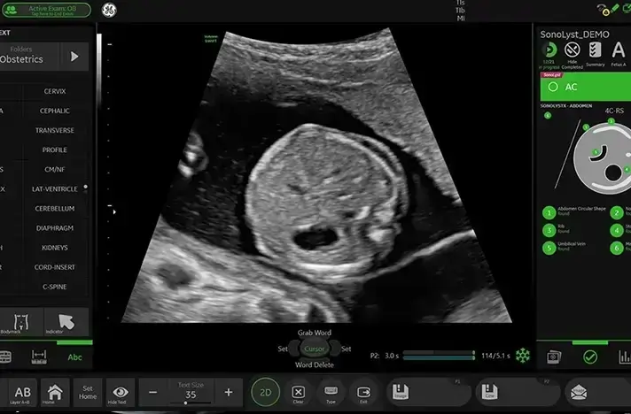
SonoLyst is the world’s first AI toolkit to automatically recognize fetal anatomical planes.
SonoLystIR: Recognizes organs, automatically annotates, and performs measurements the moment the physician freezes the image.
SonoLystlive: Operates fully automatically while the physician is scanning, helping recognize and capture standard-plane images without the physician having to perform manual operations repeatedly.
SonoCNS (Central Nervous System)
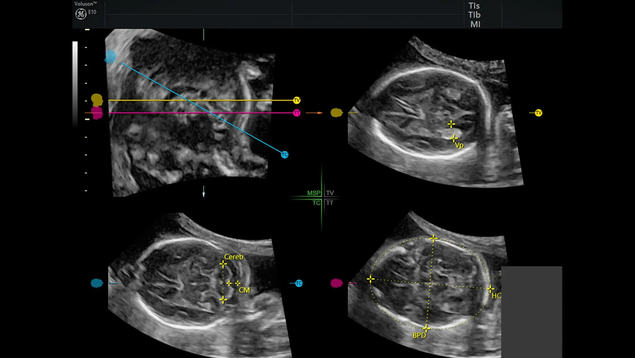
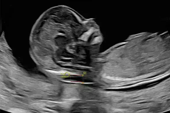
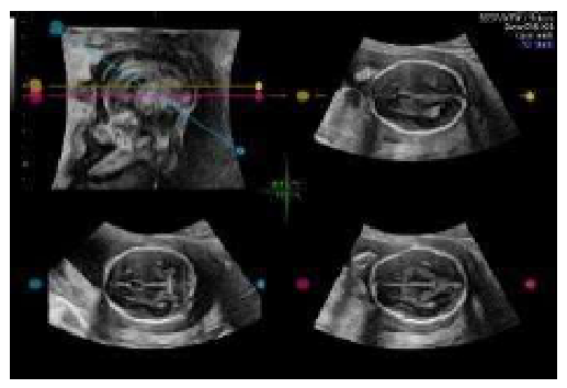
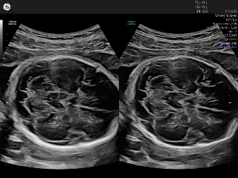
Assessing the fetal brain is one of the most difficult parts of obstetric ultrasound. SonoCNS uses deep-learning algorithms to automatically extract standard planes (the transthalamic plane, cerebellum, etc.) from a single 3D volume, and automatically performs cerebral biometric measurements. This technology helps reduce fetal brain assessment time by up to 81%.
SonoPelvicFloor
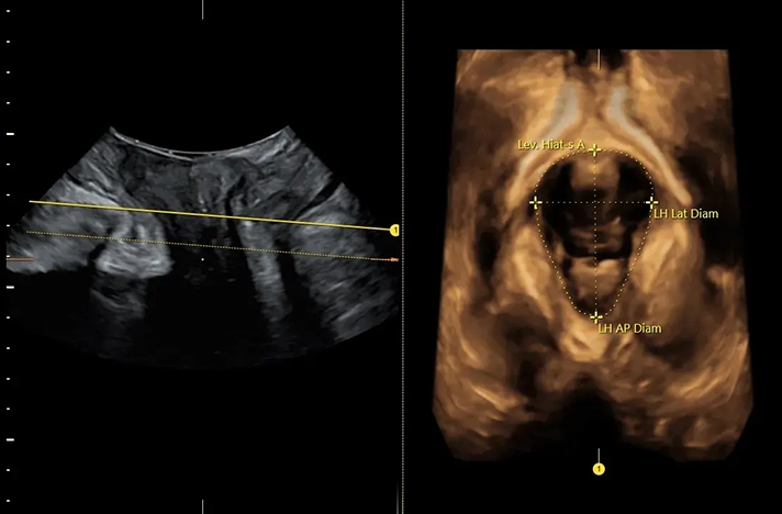
Assessing the female pelvic floor after childbirth or in cases of uterine prolapse is often complex and time-consuming. SonoPelvicFloor automates the alignment of planes and performs measurements related to the anal sphincter and bladder, helping reduce examination time by 80% and eliminating variability between different operators.
Applications In Assisted Reproductive Medicine
The Voluson line provides specialized tools that help IVF centers optimize success rates
The Voluson line provides specialized tools that help IVF centers optimize success rates.
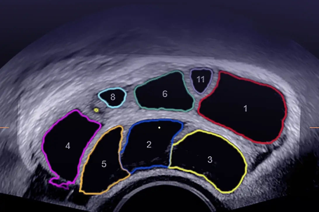
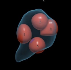
SonoAVC™follicle: Automatically counts and measures the volume of ovarian follicles. The latest version 2.0 uses AI to accurately identify follicles even when they lie close together or are surrounded by blood vessels, helping physicians make more accurate egg-retrieval decisions.
SonoAVCantral 2.0: A tool that counts antral follicles with accuracy up to 92%, supporting an objective assessment of ovarian reserve.
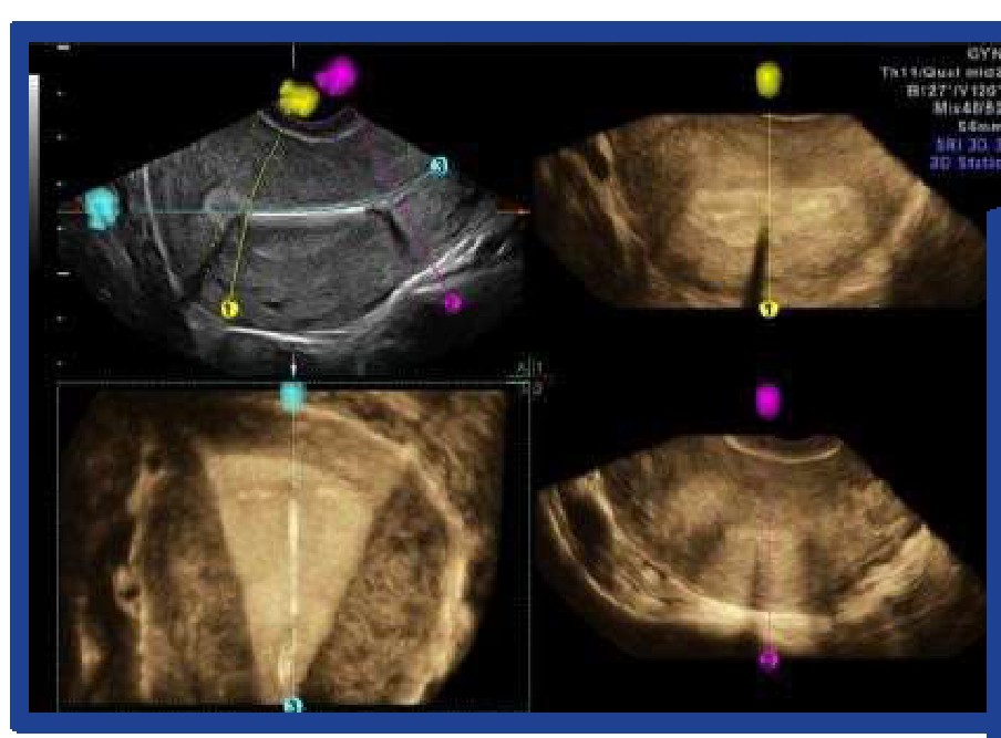
Uterine Trace: A tool that automatically reconstructs the coronal plane of the uterus in 3 simple steps, helping quickly detect uterine anomalies such as a septate uterus or heart-shaped uterus.
Ultrasound Support Software
An in-depth software suite integrated into GE HealthCare ultrasound systems — from basic image optimization to real-time AI analysis tools.
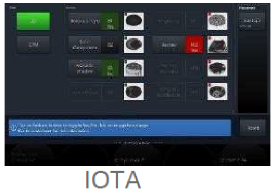
Software that supports the assessment of ovarian cysts to the international IOTA standard, with all 3 criteria: LR2, Simple Rule, ADNEX
IETA
Software that supports the survey and reporting analysis of endometrial masses
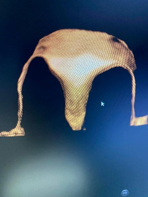
Coded Contrast Imaging + 3D HyCoSy
Software that supports ultrasound combined with contrast injection into the uterine cavity, helping reconstruct a 3D image of the fallopian tube and uterine structure, usable on the Volume Microconvex transducer
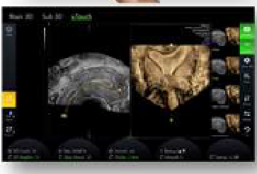
Software that creates 2D images with spatial depth
SonoLystLive (includes SonoLyst IR/SonoLystX)
Software that uses AI technology to analyze and recognize anatomical images in real time:
Automatically recognizes anatomical structures, including 20 standard planes per ISUOG guidelines, then provides suitable suggested measurements.
The ability to analyze the actual on-machine ultrasound image against the standard clinical plane, for example the presence of certain anatomical structures in the standard plane
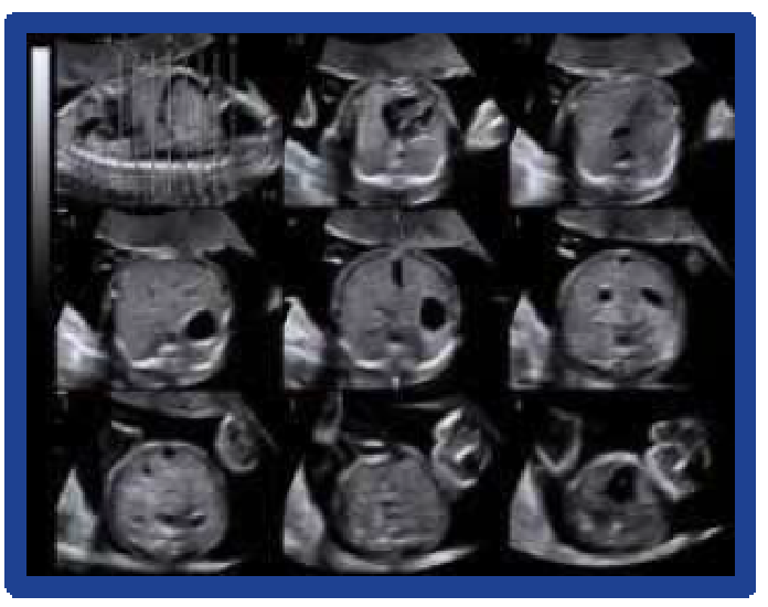
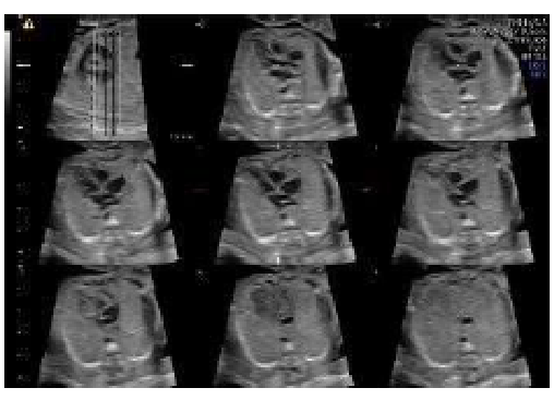
4D fetal heart and tomographic fetal heart reconstruction
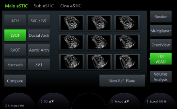
FSonoVCAD heart
Reconstruction of 7 standard planes of the fetal heart
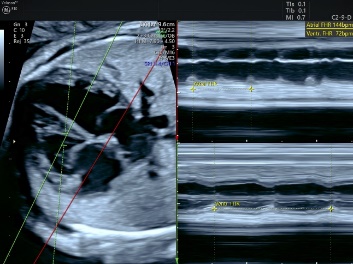
AMM
M-mode survey software that allows the M-line angle to be changed
Technology only truly matters when it translates into concrete clinical outcomes and operational efficiency. For medical facilities, an investment in imaging and AI technology should be evaluated through three lenses: diagnostic quality, productivity, and patient experience.
Earlier and more accurate diagnosis
Superior image quality together with quantification tools helps detect subtle abnormalities early — from fetal heart defects to central nervous system abnormalities. Early diagnosis opens up more intervention options and improves prognosis, while reinforcing the facility’s professional reputation.
Increasing clinic productivity
Automating repetitive steps with AI helps shorten the time per case. For high-volume facilities, even a few seconds saved per case make a significant difference in capacity over the course of a year — allowing more patients to be served without adding pressure to the team.
Enhancing the patient experience
Vivid 4D images from HDlive not only support diagnosis but also create an emotional moment of connection for expectant mothers and their families. This positive experience helps build patients’ trust in and loyalty to the medical facility.
Protecting long-term investment value
Thanks to software upgradeability, new technologies and AI tools can be added over time without replacing the hardware. This means a device purchased today can still access improvements for many years to come — an important factor when considering the total cost of ownership.
TD Tech does not just supply technology, but accompanies you so your facility can fully harness its value — through configuration advice, clinical training, and full-lifecycle technical support.
*Technology information is general in nature; specific specifications and features depend on the machine line and configuration. Please contact TD Tech for detailed advice.
Deployment & training
Technology is only powerful when the team can master it
TD Tech doesn’t stop at handing over features — we accompany you so your team can fully harness the value of the technology.
🎯
Needs assessment
Evaluating your current specialty, volume, and workflow to advise on the right technology configuration you need.
🖥️
Demo & experience
Demonstrating the technology on real patients at your facility, helping the team envision the new workflow before investing.
🧑🏫
Clinical training
Transferring the skills to harness HDlive, Edison AI, and specialized tools to physicians and technicians.
🔄
Continuous updates
Supporting software upgrades and introducing new features, keeping the equipment up to date over time.
Frequently asked questions
Answers about GE HealthCare ultrasound technology
Common questions about the imaging and artificial intelligence technologies on the Voluson line.
What is HDlive and what benefits does it bring?
HDlive is GE HealthCare’s 3D/4D rendering technology that uses a movable virtual light source to create images with rich depth and realism. It helps physicians observe subtle anatomical detail, supports diagnosis, and provides an intuitive, emotional experience for expectant mothers.
How does Edison AI support physicians?
Edison AI is an artificial intelligence platform integrating tools such as SonoLyst, SonoCNS, and SonoPelvicFloor, helping automatically recognize standard planes, take measurements, and support specialized surveys. This reduces repetitive steps, shortens time, and increases consistency across physicians.
What is the SonoAVC IVF toolkit used for?
SonoAVC is a toolkit that automatically counts and measures the volume of follicles from 3D volume data, supporting the monitoring of ovarian stimulation in assisted reproduction (IVF) in a fast, objective, and repeatable way.
Which machine lines have these technologies?
The imaging and AI technologies are available on the Voluson Expert and Signature lines depending on configuration. TD Tech will advise on a suitable configuration so your facility can properly harness the features needed for its specialty.
How do HDlive Silhouette and HDlive Flow differ from regular HDlive?
HDlive Silhouette helps you “see through” the outer tissue layers to observe the internal structure of the embryo or internal organs. HDlive Flow integrates blood-flow data into the 3D volume-rendered image, helping clearly visualize the development of the fetal vascular system — complementing HDlive Studio+, which is oriented toward realistic surface reconstruction.
What is Radiantflow?
Radiantflow uses the amplitude of the color Doppler signal to create flow images with 3D depth. This makes it easy for physicians to identify tiny blood vessels and the boundaries between them — especially important when evaluating the fetal heart and cerebral vascular system.
How does SonoLyst automatically recognize anatomical planes?
SonoLyst is the world’s first AI toolkit to automatically recognize fetal anatomical planes to international standards. SonoLystIR recognizes organs, self-annotates, and measures the moment the image is frozen; SonoLystlive operates fully automatically during scanning, helping standardize the workflow and reduce thousands of keystrokes each day.
How much faster does SonoCNS assess the fetal brain?
SonoCNS uses deep learning to automatically extract standard planes (transthalamic, cerebellum…) from a single 3D volume and automatically performs cerebral biometric measurements. This technology helps reduce fetal brain assessment time by up to 81% — one of the most difficult parts of obstetric ultrasound.
How does fetalHS support fetal heart surveys?
fetalHS (Fetal Heart Screening) automates the fetal heart survey process, helping reduce fetal heart assessment time by 48–49% while still ensuring the full set of standard planes is acquired per clinical recommendations.
What problem does Shadow Reduction solve?
Shadow Reduction automatically compensates for shadowing behind bony structures (such as the ribs or fetal spine), helping restore obscured tissue information so physicians get a more complete view with fewer missed findings.
How does the Lyric Architecture help with difficult (obese) cases?
Lyric Architecture uses graphic-based beamforming, greatly increasing raw-data processing capability to achieve superior penetration, resolution, and frame rate. This architecture is optimized for matrix transducers and AI algorithms, helping achieve accurate diagnosis even with obese patients or fetuses in unfavorable positions.
What is STIC technology used for in fetal heart assessment?
STIC (Spatio-Temporal Image Correlation) acquires a dynamic volume of the fetal heart and reconstructs it according to the cardiac cycle, allowing the fetal heart to be examined in 3D/4D. This technique helps analyze the structure and function of the fetal heart in more detail than conventional 2D.