 Email: tdmedical@gmail.com



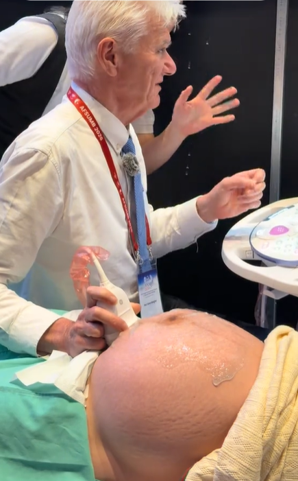
Professor Simon Meagher and Professor Ritsuko Pooh Performing Ultrasound Examinations Using the Voluson E22 System
 8.3K
8.3K
 51
51
 09-Aug-2026
 61,845

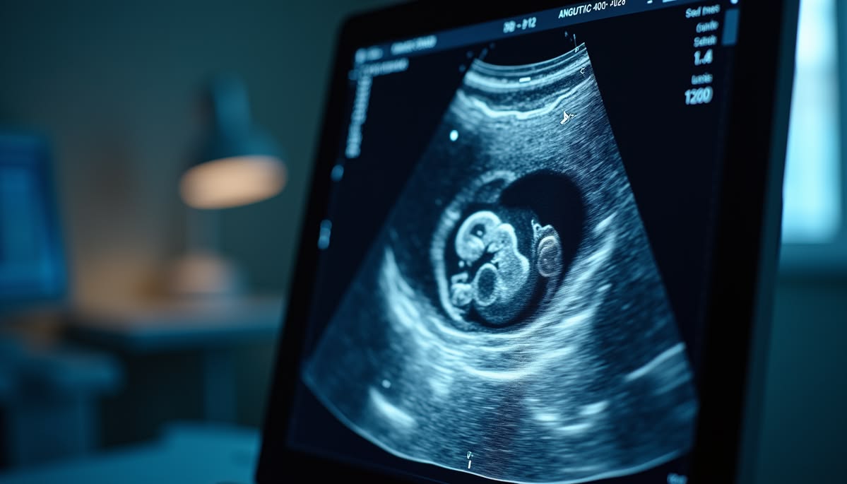
2D ultrasound is a technique that produces real-time, two-dimensional cross-sectional images of structures inside the body. The transducer emits ultrasound waves into the body and receives reflected signals from tissues. The system then processes these signals to generate images on the screen.

This is the fundamental ultrasound imaging method and is widely used across various specialties, including obstetrics and gynecology, cardiology, abdominal imaging, thyroid imaging, vascular imaging, and musculoskeletal imaging.
With 2D ultrasound, physicians can observe anatomical structures, perform measurements of distance, size, and area, and combine imaging with Doppler modes to assess blood flow and hemodynamics.
However, 2D ultrasound displays individual cross-sectional images. Therefore, visualizing the spatial relationships between complex structures may depend considerably on the skill and experience of the operator.
3D ultrasound builds upon conventional 2D imaging by enabling the acquisition of a volumetric dataset of the region being examined.
In simple terms, the process can be described as follows:
Image data acquisition → volumetric dataset generation → processing and three-dimensional image reconstruction.
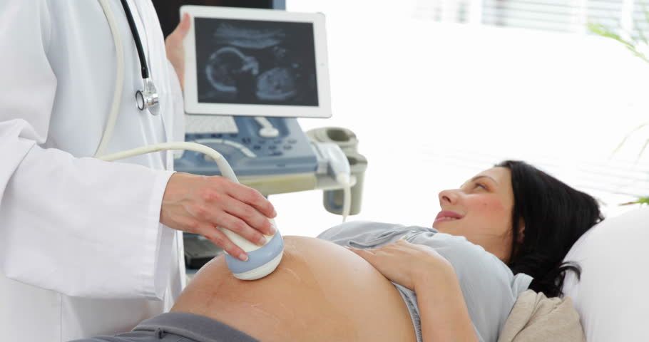
From the acquired volume, the system can allow physicians to examine structures in multiple planes, including certain planes that may be difficult to obtain directly with conventional 2D ultrasound.
Importantly, 3D data is not limited to creating more intuitive images. In appropriate applications, the volume dataset can also be stored and reanalyzed after acquisition, supporting a more systematic assessment of anatomical structures.
While 3D ultrasound provides volumetric data of a structure at a specific point in time, 4D ultrasound adds the dimension of time by continuously updating volumetric datasets.
Simply put:
3D = three-dimensional volumetric data.
4D = three-dimensional volumetric data changing over time.
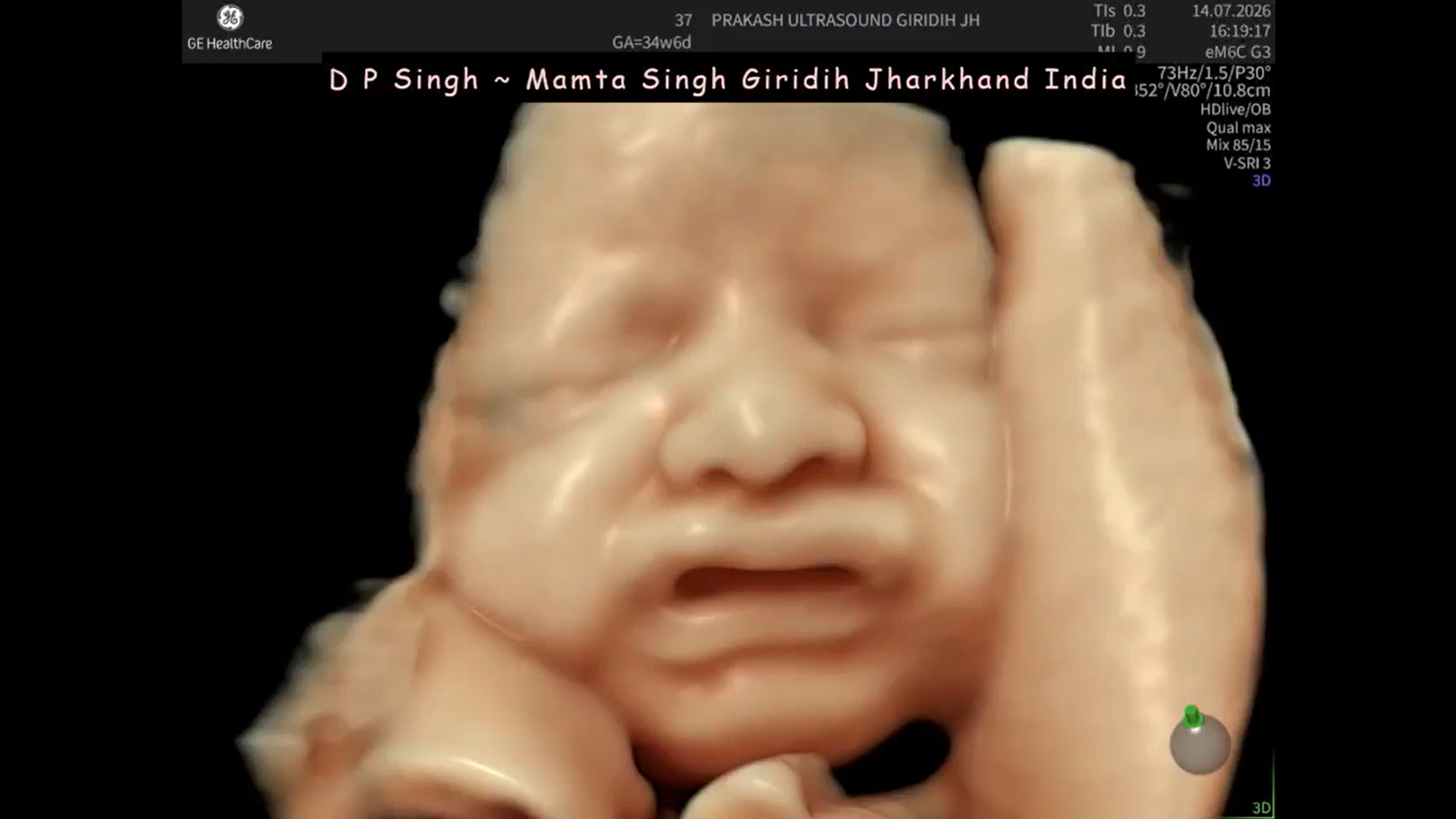
As a result, 4D ultrasound enables structures and their movements to be observed as dynamic volumetric images. In obstetrics, this technology is commonly known for visualizing fetal movements, facial features, and limbs. However, the value of 4D ultrasound is not limited to producing intuitive fetal images; depending on the system, it can also be used in selected advanced diagnostic applications.
| Criteria | 2D Ultrasound | 3D Ultrasound | 4D Ultrasound |
|---|---|---|---|
| Data type | Two-dimensional cross-sectional data | Three-dimensional volumetric data | Volumetric data over time |
| Display | Cross-sectional images | 3D images/volumes | Dynamic 3D images |
| Multiplanar examination | More limited | Yes | Yes |
| Volume analysis | Not a primary function | Yes | Yes |
| Motion visualization | Available with real-time 2D imaging | Not the primary purpose | Available as dynamic volumetric imaging |
| Data processing | Simpler | More complex | More complex |
| Applications | Very broad | Selected clinical indications | Selected clinical indications |
An important point is that 3D/4D ultrasound was not developed to completely replace 2D ultrasound. In many workflows, physicians use 2D ultrasound for the initial examination and add 3D/4D when additional information about spatial anatomy or volumetric data is required.
One advantage of volumetric data is the ability to examine structures from multiple planes. This can help physicians better visualize spatial relationships between anatomical structures, particularly when a single 2D cross-sectional image does not provide all the necessary information.
3D data enables volumetric analysis of certain structures and lesions. Depending on the software, physicians may also be able to manipulate, rotate, crop, or re-examine the volume after the dataset has been acquired.
This capability is particularly useful in fields where complex spatial anatomy needs to be evaluated, such as obstetrics and gynecology.
Surface and volume rendering techniques can make certain anatomical structures easier to visualize. In addition to supporting physicians in appropriate clinical indications, 3D/4D images may also help explain examination findings to patients more clearly.
Obstetrics is one of the most common fields in which 3D/4D ultrasound technology is applied.

3D imaging can provide additional information when examining structures such as the fetal face, limbs, spine, and body surface. In appropriate cases, spatial reconstruction can help physicians assess the morphological characteristics of the structures being examined more clearly.
Acquisition of fetal brain volumes allows data to be examined in multiple planes from the same dataset. This can serve as an additional tool when assessing certain central nervous system structures in cases requiring advanced evaluation.
Volumetric technologies can support the acquisition and analysis of data related to fetal cardiac structures. However, 3D/4D ultrasound is a complementary tool and does not replace specialized fetal echocardiography when clinically indicated.
4D ultrasound can display movements of the fetal face, hands, legs, and other body parts as dynamic volumetric images. This is also one of the features that has made 4D ultrasound widely recognized among patients during pregnancy monitoring.
Beyond obstetrics, volumetric imaging also has various applications in gynecologic ultrasound.
One notable advantage of 3D ultrasound is its ability to reconstruct the coronal plane of the uterus, a plane that can be difficult to obtain directly with conventional 2D ultrasound. This view may provide additional information when assessing uterine morphology and certain structural abnormalities.
3D data can also be used to examine the endometrium, uterine cavity, ovaries, and adnexal structures. Depending on the clinical situation, volumetric analysis and Doppler tools may be combined to provide additional information for physicians.
It cannot be stated that 3D/4D ultrasound is always more accurate than 2D ultrasound.
Diagnostic value depends on many factors, including the organ being examined, clinical indication, quality of the initial images, ultrasound system, transducer, processing software, and the operator’s experience.
In many cases, 2D ultrasound provides sufficient diagnostic information and remains the primary examination method. Conversely, for structures or indications requiring spatial and volumetric assessment, 3D/4D ultrasound can provide additional information that may be difficult to obtain from individual 2D planes.
Therefore, rather than viewing 2D and 3D/4D ultrasound as competing technologies, they should be considered complementary imaging methods within the diagnostic workflow.
2D ultrasound is suitable for most routine examinations, ranging from pregnancy monitoring to cardiac, abdominal, thyroid, and vascular ultrasound. It is particularly useful when physicians need to rapidly examine cross-sectional views and observe structures in real time.
3D/4D ultrasound may be added when there is a need to:
The choice of imaging method should be based on clinical indications and examination objectives rather than solely on how advanced the technology is.
Next-generation ultrasound systems increasingly combine volumetric acquisition technologies with advanced image-processing algorithms and automation tools to optimize examination workflows.
In obstetrics and gynecology, these systems may provide volume rendering, surface rendering, multiplanar display, and volumetric data analysis. Some platforms also integrate tools that support automatic structure recognition, standardized plane acquisition, or automated measurements, depending on the ultrasound system and installed software package.
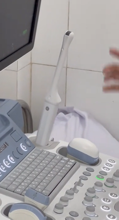
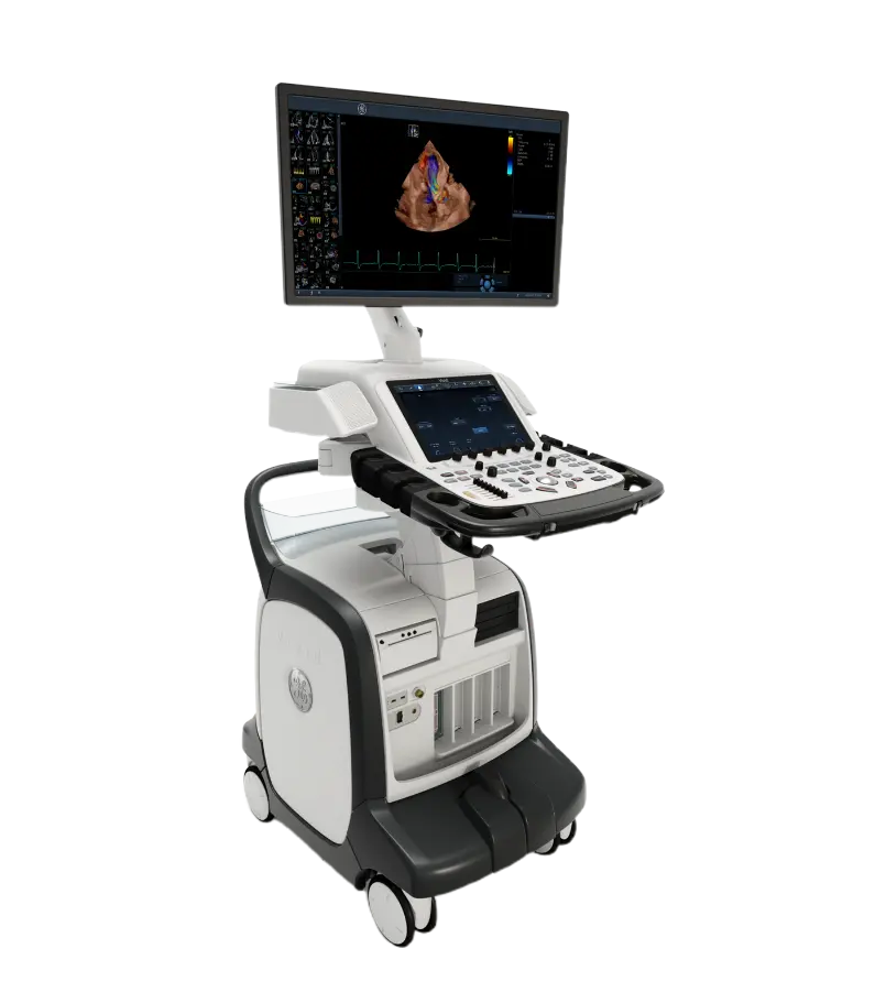
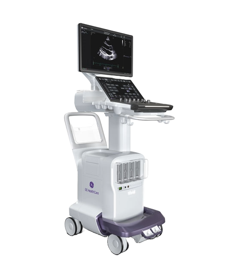
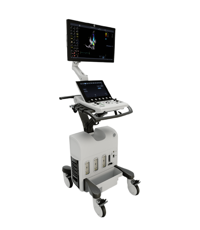
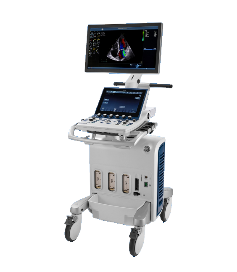
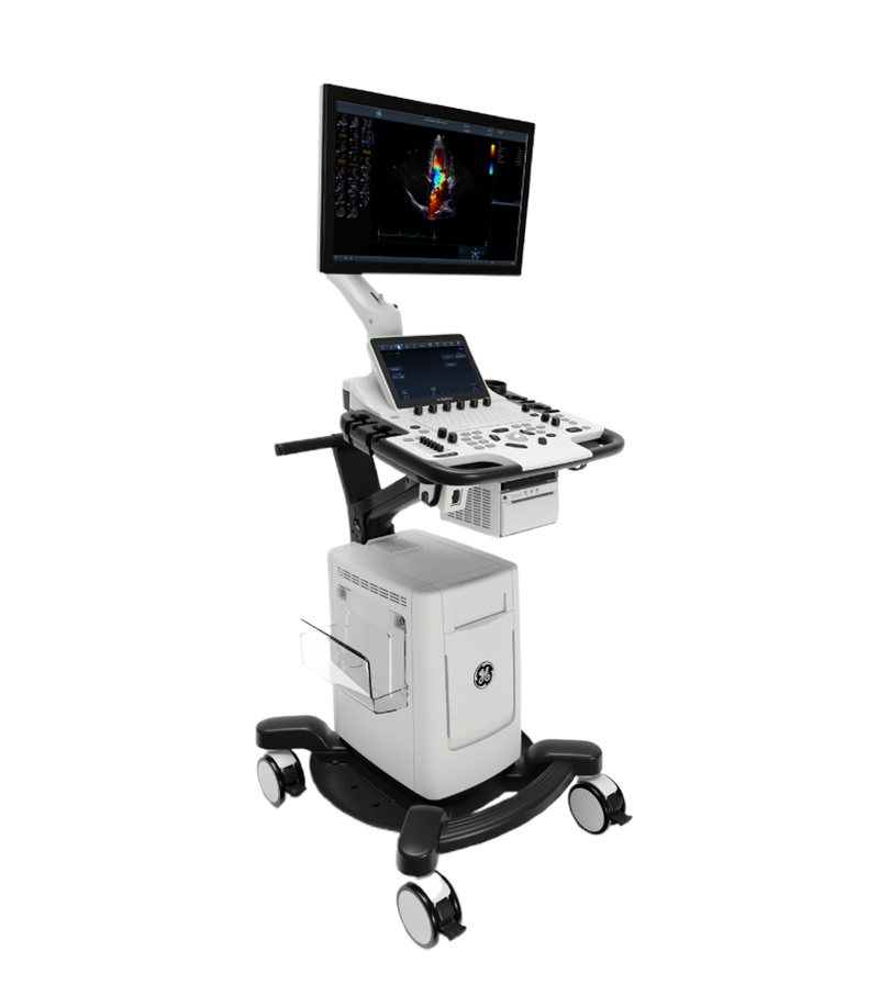
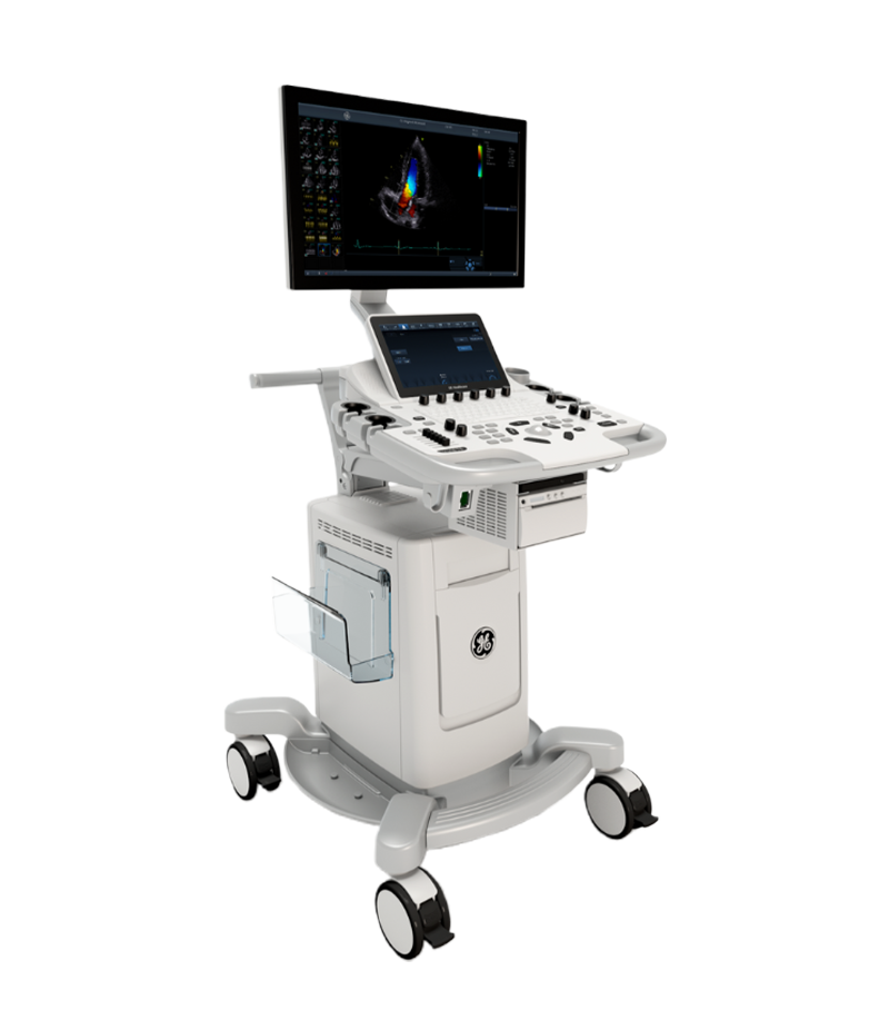
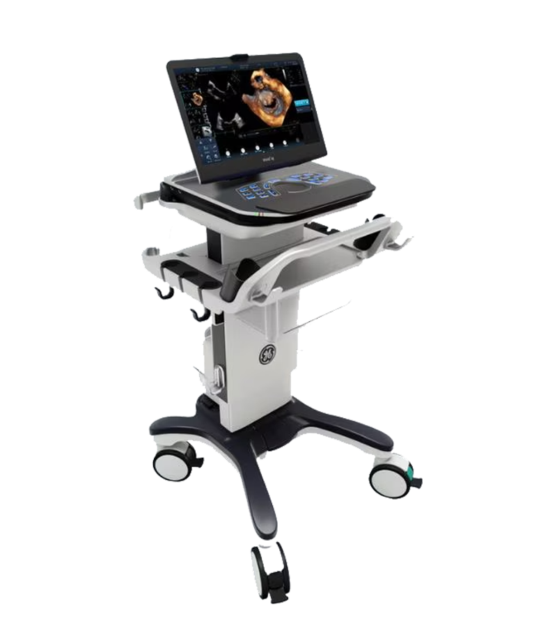
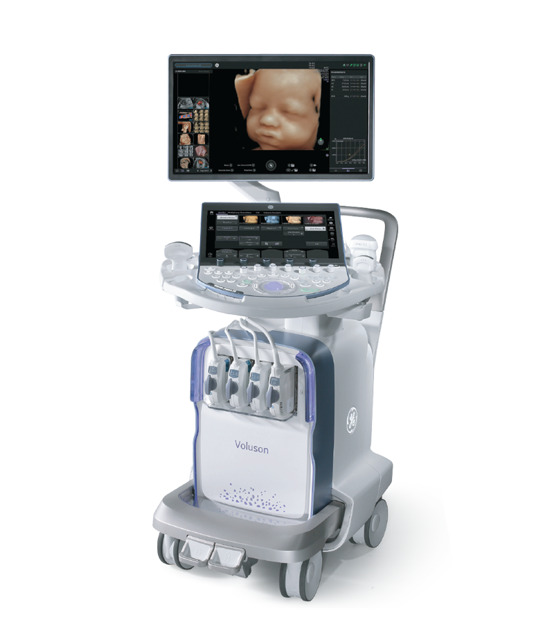
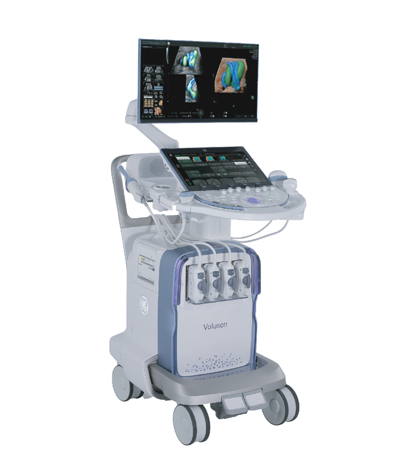
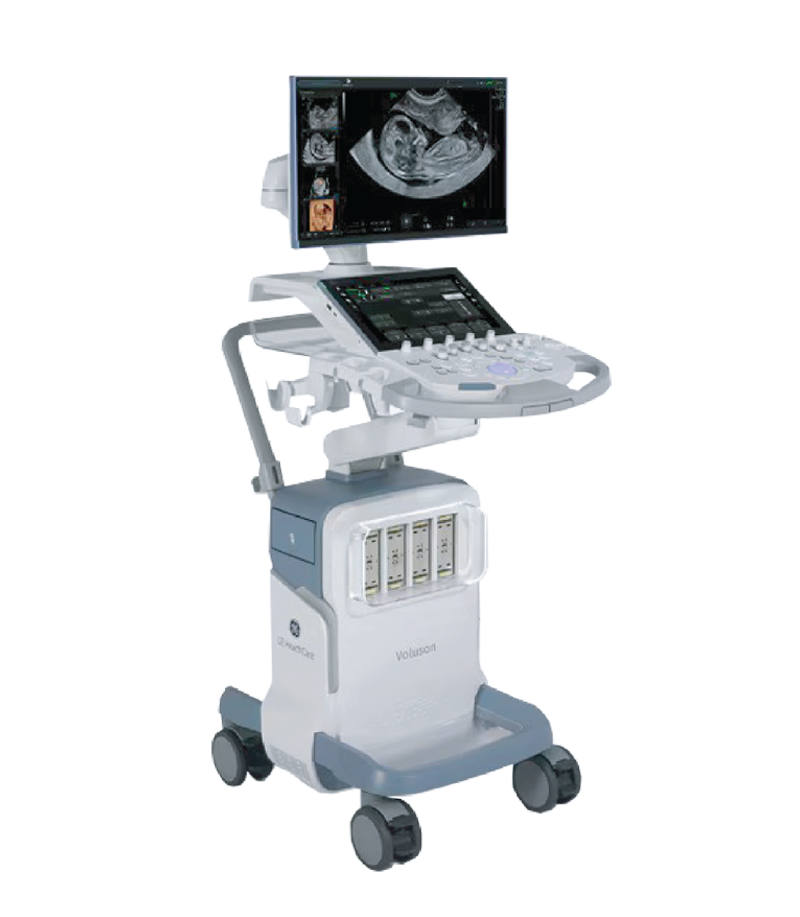
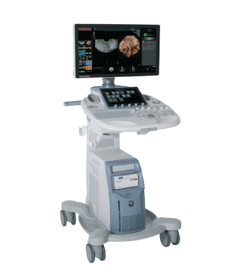
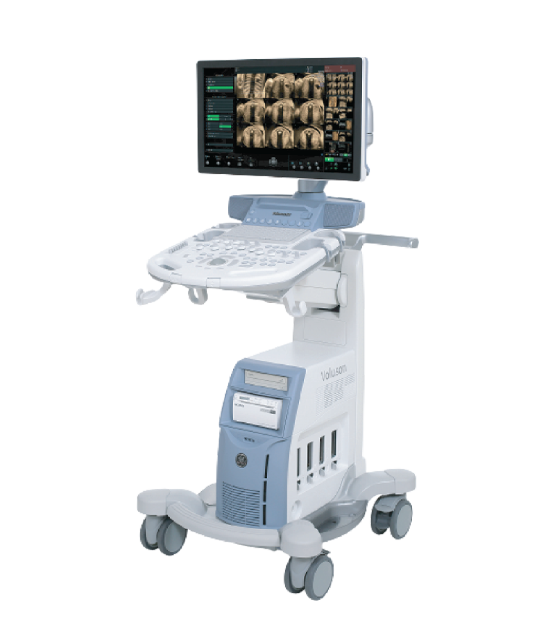
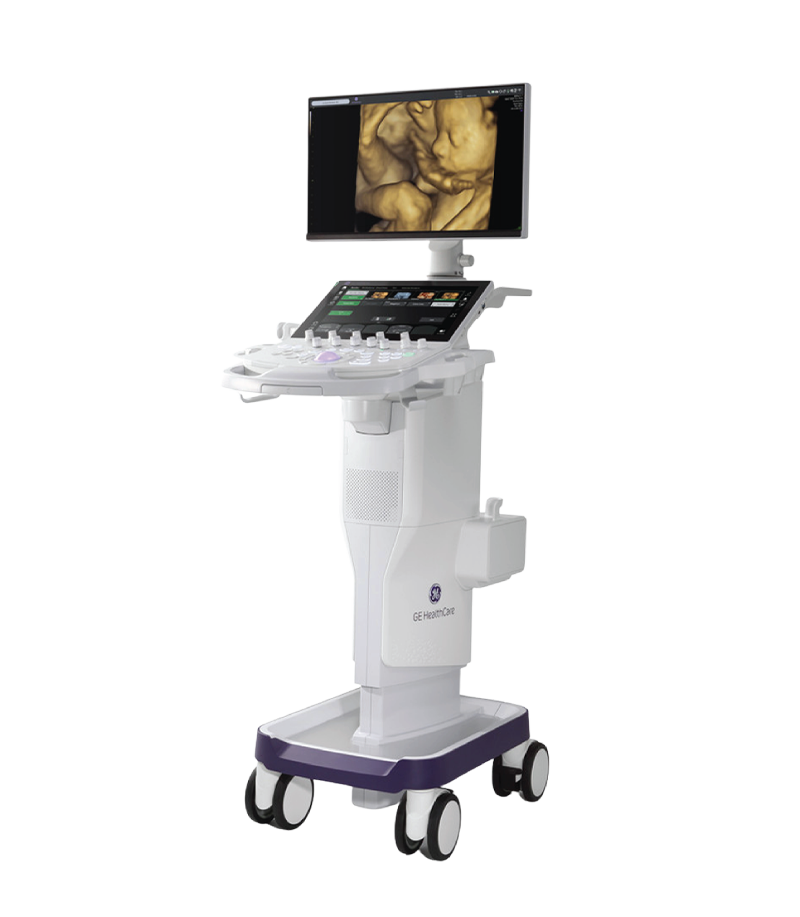
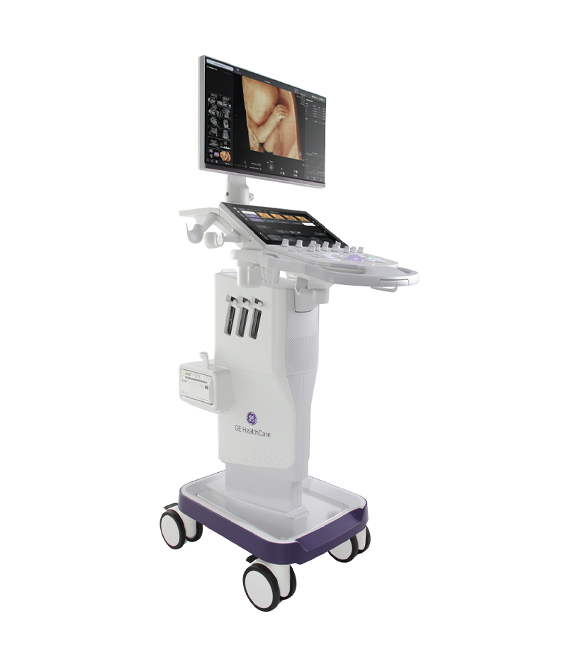
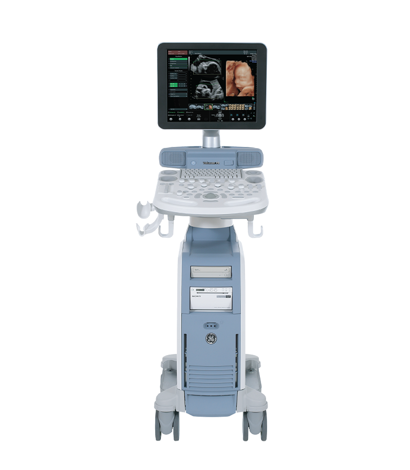
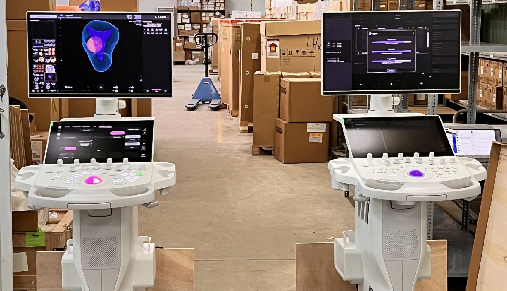
As a provider of GE HealthCare ultrasound solutions, TD TECH offers Voluson obstetric and gynecologic ultrasound systems with a range of configurations supporting 3D/4D technology and advanced image-processing tools. Depending on clinical needs, from routine examinations to advanced diagnostic imaging, healthcare facilities can select the appropriate system configuration, transducers, and software to suit their professional workflows.
Cardiac and Obstetric Ultrasound Systems
The appropriate integration of 2D, 3D/4D, Doppler, and modern image-processing technologies can help physicians obtain more comprehensive ultrasound information while optimizing examination and diagnostic workflows.
“No crying, no thinking… but definitely very expressive.” At 34–35 weeks of pregnancy, a fetus’s facial expressions can change within just a few seconds. From one ultrasound frame to the next, it can sometimes...
Xem thêm
Throughout pregnancy, the moment parents see their baby’s face and movements in the womb is always one of the most precious and emotional experiences. Thanks to remarkable advances in modern medicine, 3D/4D ultrasound has become...
Xem thêm
What Is Twin-to-Twin Transfusion Syndrome? Twin-to-Twin Transfusion Syndrome (TTTS) is a serious complication that can occur in monochorionic twin pregnancies, in which vascular anastomoses within the shared placenta lead to an imbalanced exchange of blood...
Xem thêm
Every time expectant parents receive an ultrasound report, they are often confronted with a long list of unfamiliar abbreviations such as BPD, FL, CRL, NT, and AFI. To support families throughout pregnancy, TD TECH has...
Xem thêm
Unboxing the Voluson P16 and P18 ultrasound machines – the two latest models in the P-Series featuring modern designs, sharp imagery, and effective support for OB/GYN and multi-specialty diagnostics. Introduction In the field of diagnostic...
Xem thêm
What Is a Fetal Anatomy Ultrasound? A fetal anatomy ultrasound is an ultrasound examination designed to systematically assess the anatomical structures and development of the fetus. During the examination, the physician evaluates the fetus using...
Xem thêm





 Xem video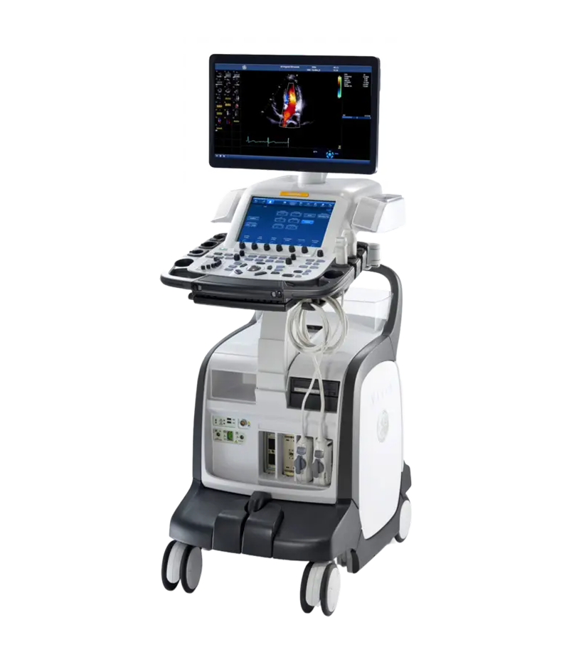





 Consult Now