 Email: tdmedical@gmail.com




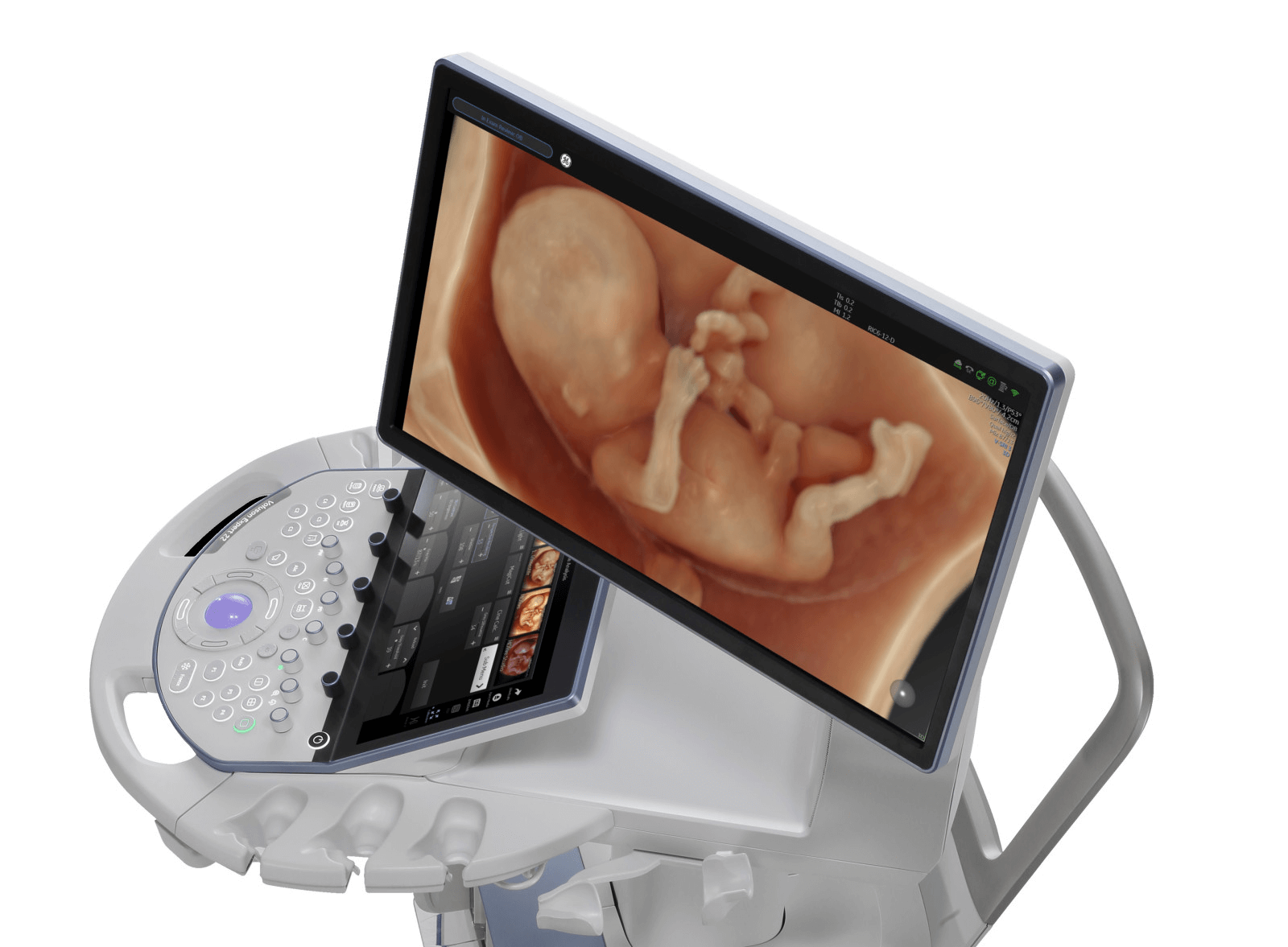
Professor Simon Meagher and Professor Ritsuko Pooh Performing Ultrasound Examinations Using the Voluson E22 System
 8.3K
8.3K
 51
51
 18-Aug-2026
 81,700

A fetal anatomy ultrasound is an ultrasound examination designed to systematically assess the anatomical structures and development of the fetus.
During the examination, the physician evaluates the fetus using multiple standardized imaging planes to assess important organs and body structures. Fetal biometric measurements are also obtained to determine whether fetal size is appropriate for gestational age.
A fetal anatomy assessment generally includes four main groups of information:
The primary goal is to identify or raise suspicion of structural abnormalities and determine which pregnancies may require more detailed evaluation.
A detailed fetal anatomy scan is usually performed during the second trimester, commonly between 18 and 22 weeks of gestation according to many international guidelines. In clinical practice, the timing may vary between healthcare facilities, for example between 20 and 24 weeks, depending on local protocols.

In addition to the mid-trimester anatomy scan, fetal anatomical assessment may also be performed at other stages of pregnancy. Some abnormalities can be identified during the first trimester, while others may become apparent or progress only later in pregnancy.
A fetal anatomy scan does more than provide an overall image of the fetus. The physician systematically examines multiple structures using standardized imaging planes.
The physician evaluates the shape and integrity of the skull as well as the intracranial structures that can be visualized at the corresponding gestational age.
The examination commonly includes structures such as the midline of the brain, lateral ventricles, thalami, posterior fossa, and cerebellum on appropriate imaging planes. Head measurements such as biparietal diameter and head circumference also provide important information about fetal growth.
If suspicious findings are detected, a detailed fetal neurosonographic examination may be recommended for further assessment.
During a fetal anatomy ultrasound, the face is examined in multiple planes to assess structures such as the orbits, lips, and facial profile. Certain facial abnormalities, particularly some forms of cleft lip, may be detected by ultrasound. However, visualization depends significantly on fetal position, amniotic fluid volume, and overall imaging conditions.
When necessary, 3D ultrasound may provide additional surface views, but it does not replace the standard 2D anatomical examination.
The fetal spine is examined in multiple planes to assess its continuity, shape, and relationship with the overlying soft tissues. The goal is to identify findings that may suggest abnormalities such as open neural tube defects or certain vertebral structural abnormalities.
The physician generally evaluates not only the spine itself but also associated findings in the brain and skull that may be related to neural tube abnormalities.
The heart is one of the most important and complex components of the fetal anatomy examination.
Fetal cardiac assessment goes beyond simply confirming fetal cardiac activity or viewing the four chambers. The physician may also evaluate cardiac position and axis, heart rhythm, the four-chamber view, ventricular outflow tracts, great vessels, and additional cardiac views according to professional examination protocols.
The goal is to identify congenital heart abnormalities that may be detectable on prenatal ultrasound. If suspicious findings, risk factors, or incomplete cardiac visualization are present, the physician may recommend a detailed fetal echocardiography examination.
The physician evaluates the shape of the fetal chest as well as the position of the heart, lungs, and diaphragm. The relationship between these organs is also assessed to identify abnormalities that may alter normal thoracic anatomy.
Certain conditions, such as congenital diaphragmatic hernia or space-occupying lesions within the chest, may displace the heart or other organs.
The abdominal wall is examined to assess its integrity and the site where the umbilical cord enters the fetal abdomen. The physician also evaluates the presence and position of the stomach. Some abnormalities of the abdominal wall or gastrointestinal tract may present as abnormal morphology, dilated structures, or changes in amniotic fluid volume.
However, not all gastrointestinal abnormalities can be identified during the mid-trimester fetal anatomy scan.
Both kidneys, their location, and the fetal bladder are examined to assess the urinary system. Findings that may receive particular attention include dilation of the urinary collecting system, abnormal renal morphology, an abnormal bladder, or failure to visualize structures that would normally be expected.
The fetal urinary system is also closely related to amniotic fluid volume, particularly during later stages of pregnancy.
The physician checks for the presence of all four limbs, evaluates the different limb segments, and assesses the morphology of the long bones. Femur length is one of the standard fetal biometric measurements. Depending on the examination protocol and clinical indication, other long bones may also be assessed.
Fetal anatomy ultrasound is considered very safe for both the mother and the fetus. Ultrasound uses high-frequency sound waves to generate images and does not use ionizing radiation such as X-rays.
A normal umbilical cord usually contains three blood vessels: two umbilical arteries and one umbilical vein.
During the ultrasound examination, the physician may assess the number of vessels, the site where the umbilical cord enters the fetal abdominal wall, and the placental cord insertion, depending on visualization and the examination protocol.
If an umbilical cord abnormality is identified, its clinical significance should be interpreted together with fetal anatomy, growth, and other relevant parameters.
Amniotic fluid volume is also assessed during the examination. Oligohydramnios or polyhydramnios may be associated with a variety of maternal, fetal, or placental conditions. Amniotic fluid volume also directly affects how well the fetus can be visualized during ultrasound.
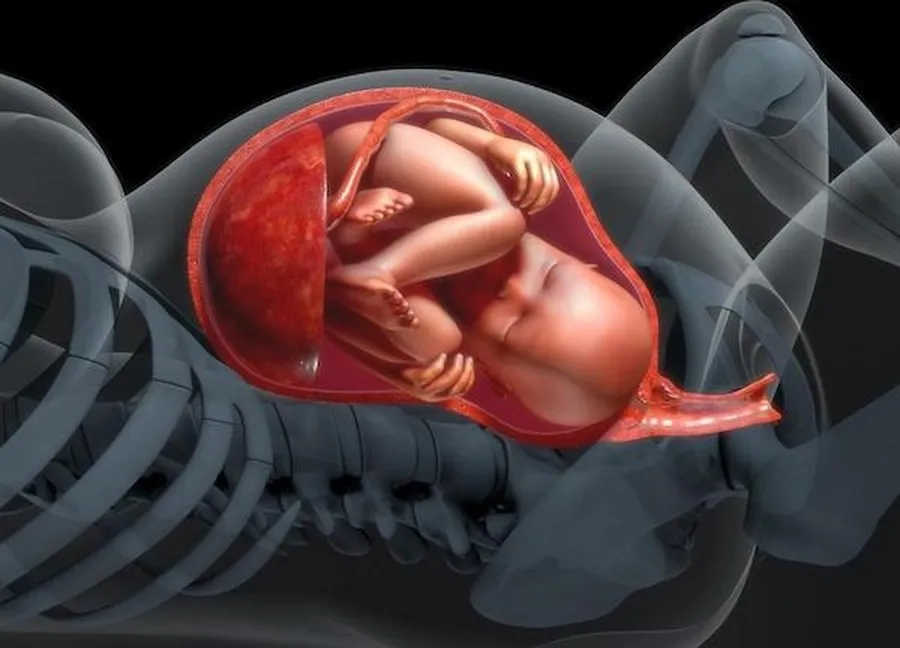
Unlike some commonly used descriptions, ultrasound cannot determine the “color” of amniotic fluid. Ultrasound primarily helps assess amniotic fluid volume and certain related imaging characteristics.
Doppler ultrasound does not necessarily need to be used in the same way for every pregnancy, but it can provide important hemodynamic information when clinically indicated. Depending on gestational age and the clinical situation, the physician may evaluate:
Uterine arteries: provide information about uteroplacental circulation and may be used to assess the risk of certain placenta-related pregnancy complications.
Umbilical artery: reflects resistance within the placental circulation and is particularly important when monitoring pregnancies affected by fetal growth problems.
Middle cerebral artery (MCA): has several specialized applications, including assessing fetal circulatory redistribution or peak systolic velocity in specific clinical indications.
Ductus venosus (DV): provides additional information about fetal venous hemodynamics and cardiovascular function in selected high-risk situations.
In summary, fetal anatomy ultrasound focuses primarily on anatomical structures, while Doppler provides additional information about blood flow and fetal hemodynamics when clinically indicated.
No. This is an important limitation that pregnant women should understand before undergoing the examination.
The ability to detect abnormalities depends on many factors, including whether the abnormality has become apparent at the time of the ultrasound examination.
Some congenital abnormalities are clearly visible on ultrasound, whereas small lesions or functional disorders may be difficult to detect. In addition, certain conditions may only become apparent later in pregnancy or after birth.
Therefore, a normal fetal anatomy ultrasound reduces the likelihood of many detectable structural abnormalities but cannot guarantee that the fetus is completely free of abnormalities.
A fetal anatomy ultrasound is not a diagnostic test for chromosomal abnormalities.
Certain structural abnormalities or ultrasound markers may be associated with an increased risk of chromosomal abnormalities. When such findings are identified, the physician will interpret them together with maternal age, medical and obstetric history, prenatal screening results, and other risk factors.
Depending on the individual case, additional counseling may be provided regarding prenatal screening tests, NIPT, amniocentesis, or more advanced genetic testing.
It is not always possible to obtain all the required imaging planes during a single examination.
For example, fetal position may make it difficult to visualize the heart, face, or spine. Imaging conditions may also be affected by amniotic fluid volume, the maternal abdominal wall, or placental position.
In such situations, the physician may schedule a follow-up ultrasound to complete the assessment of structures that could not be adequately visualized during the initial examination.
The quality of a fetal anatomy examination depends primarily on the examiner’s expertise, the examination protocol, and the imaging capabilities of the ultrasound system.
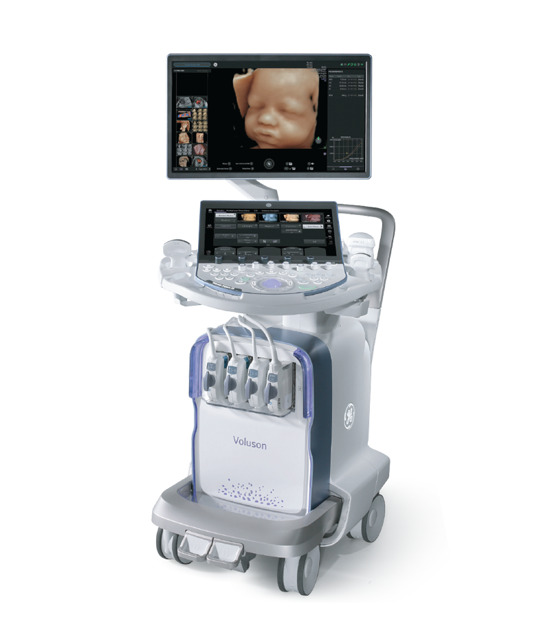
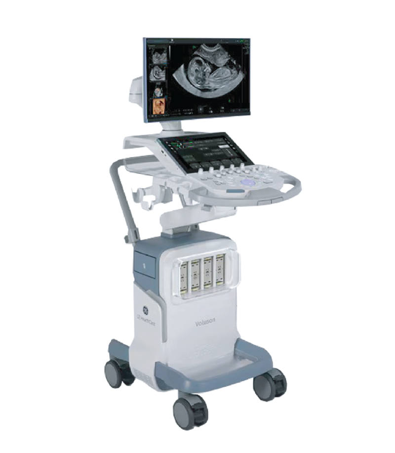
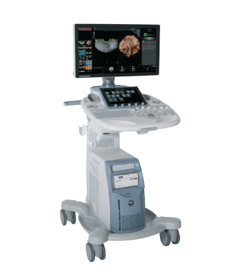
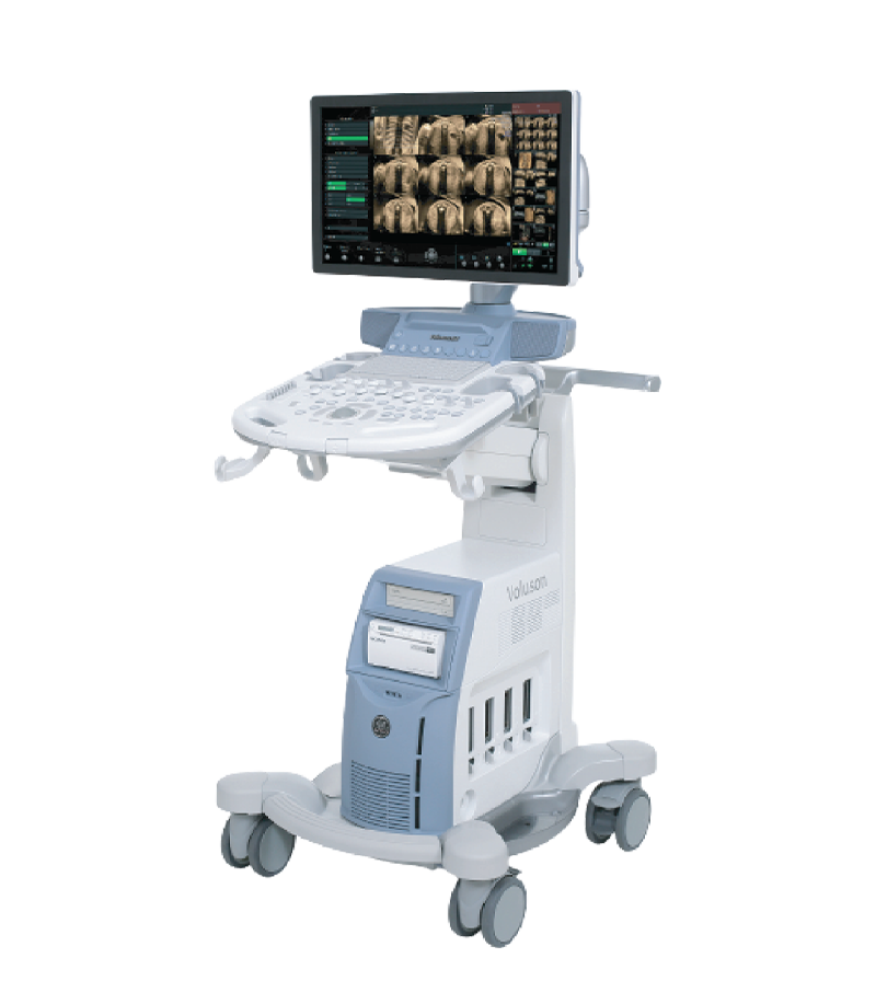
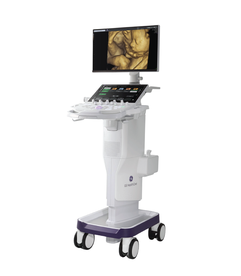
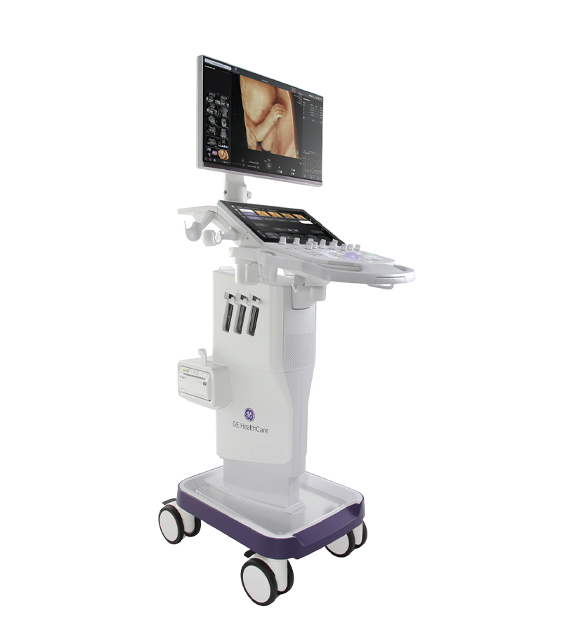
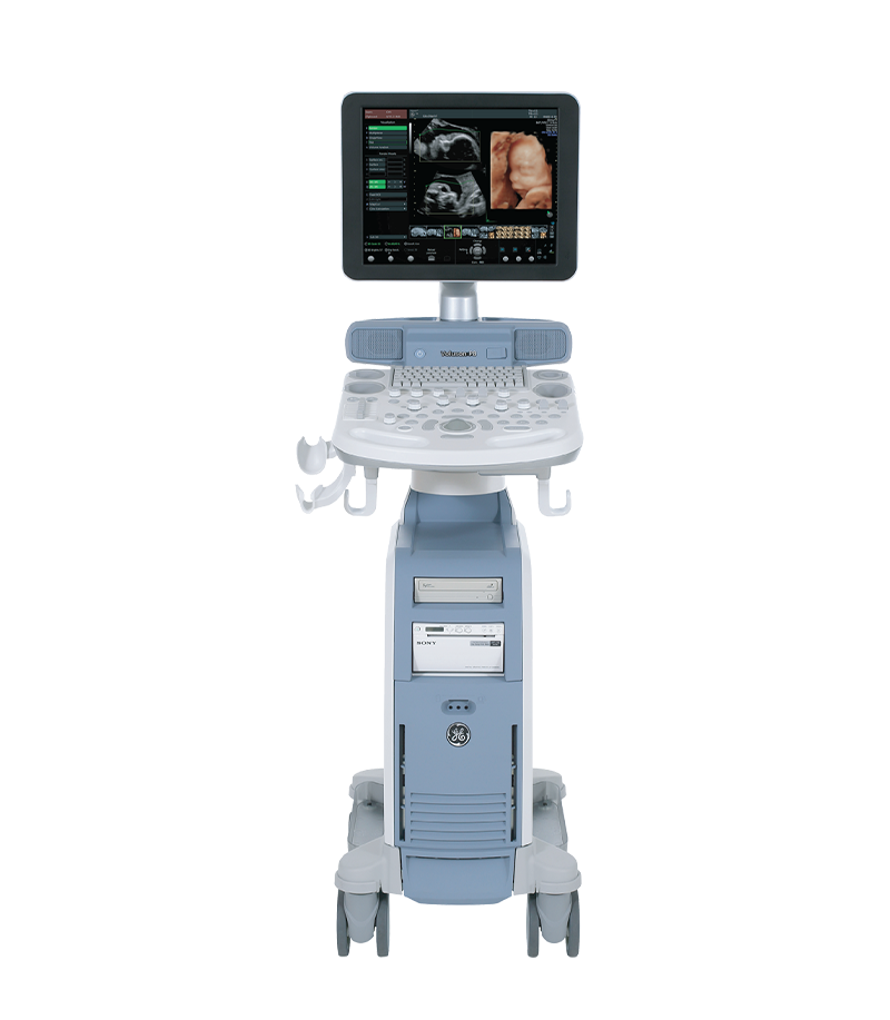
Modern obstetric and gynecologic ultrasound systems, such as Voluson, increasingly provide a wide range of supporting technologies, including high-resolution 2D imaging, color Doppler, pulsed-wave Doppler, 3D/4D volume imaging, multiplanar imaging, and automated measurement tools. The choice of an ultrasound system should be based on clinical needs, transducer options, image quality, Doppler capabilities, 3D/4D technology, and the workflow requirements of each healthcare facility.
Voluson Ultrasound System
GE HealthCare is a global medical technology company operating across diagnostic imaging, ultrasound, patient monitoring, diagnostic pharmaceuticals, AI, and healthcare software solutions. GE HealthCare offers a broad portfolio of ultrasound systems, with each product family...
Xem thêm
Throughout pregnancy, the moment parents see their baby’s face and movements in the womb is always one of the most precious and emotional experiences. Thanks to remarkable advances in modern medicine, 3D/4D ultrasound has become...
Xem thêm
What Is Valvular Heart Disease? Common Types of Valve Disorders Valvular heart disease is a condition in which one or more of the heart valves (the mitral valve, tricuspid valve, aortic valve, or pulmonary valve)...
Xem thêm
The emergence of diagnostic support software for Doppler ultrasound integrated with artificial intelligence (AI) has marked a major breakthrough, helping automate measurements, improve accuracy, and optimize workflow efficiency for physicians. What Is Doppler Ultrasound and...
Xem thêm
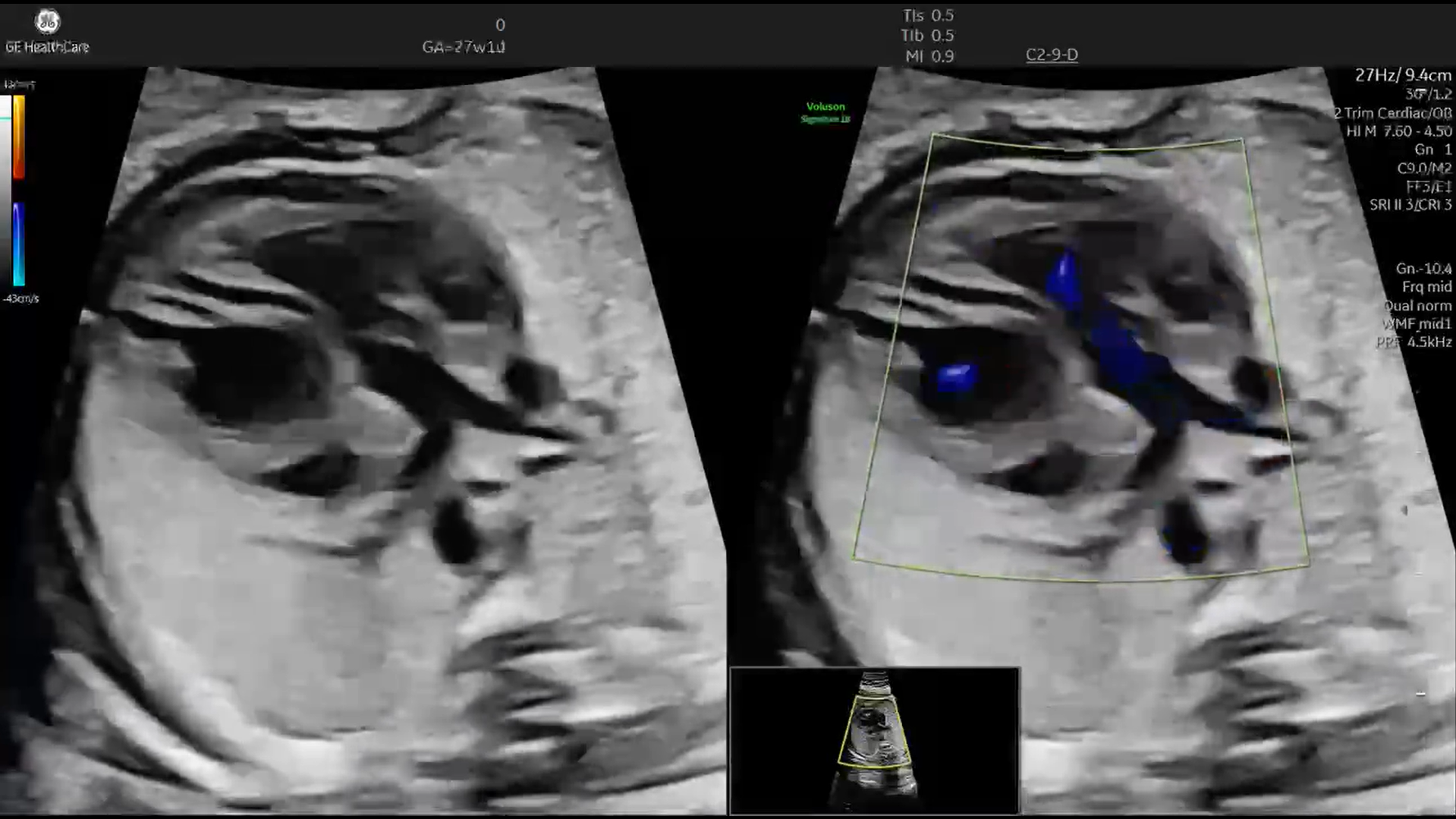
A 27-week-old fetus was referred for further evaluation after an abnormal orientation of the great arteries was detected during a routine ultrasound examination. What initially appeared to be only a subtle anatomical variation quickly...
Xem thêm
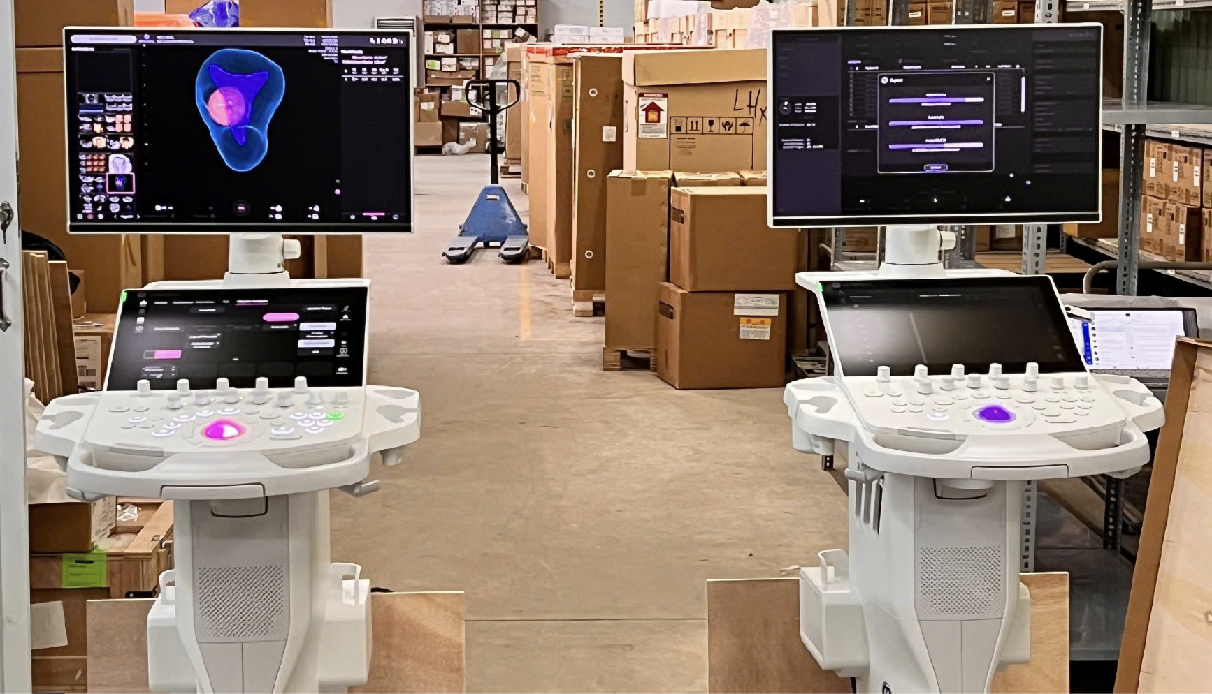
Unboxing the Voluson P16 and P18 ultrasound machines – the two latest models in the P-Series featuring modern designs, sharp imagery, and effective support for OB/GYN and multi-specialty diagnostics. Introduction In the field of diagnostic...
Xem thêm


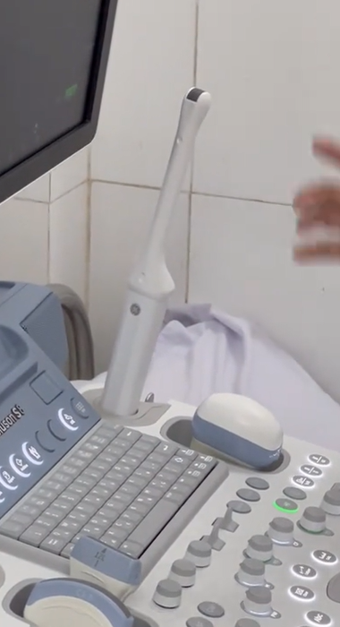


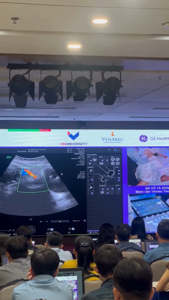
 Xem video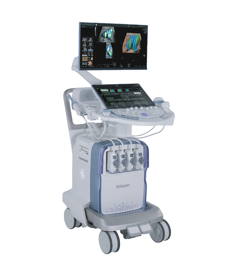





 Consult Now