 Email: tdmedical@gmail.com



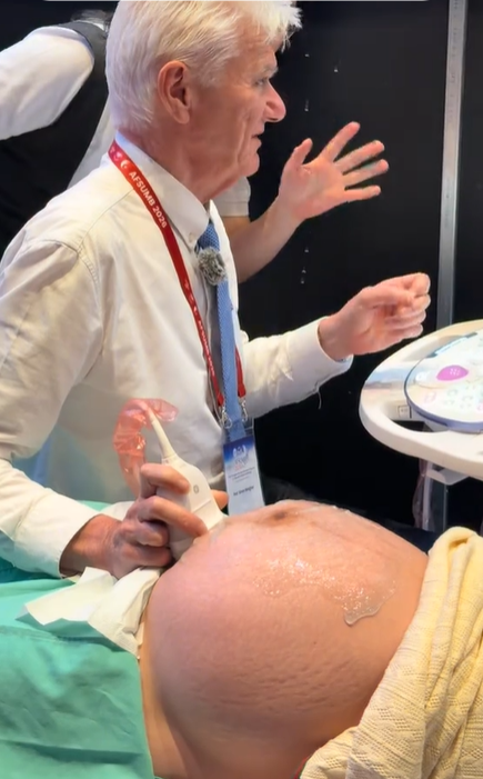
Professor Simon Meagher and Professor Ritsuko Pooh Performing Ultrasound Examinations Using the Voluson E22 System
 8.3K
8.3K
 51
51
 06-Aug-2026
 62,700

The emergence of diagnostic support software for Doppler ultrasound integrated with artificial intelligence (AI) has marked a major breakthrough, helping automate measurements, improve accuracy, and optimize workflow efficiency for physicians.
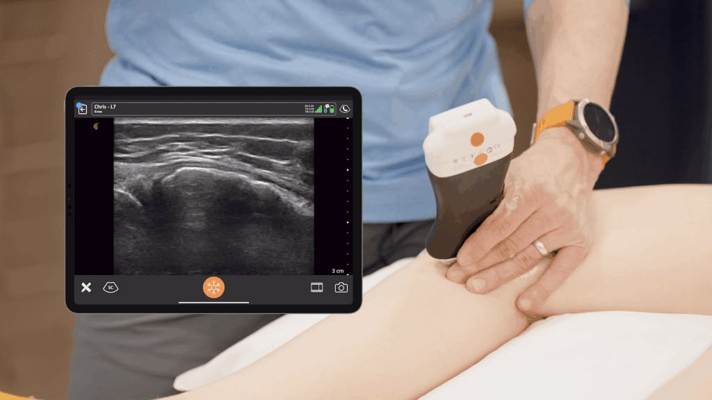
Doppler ultrasound is an ultrasound technique that uses the Doppler effect to assess the movement of blood flow within the heart and vascular system. By analyzing changes in the frequency of ultrasound waves reflected from moving blood cells, the system can provide information about blood flow direction, velocity, and hemodynamic characteristics in the area being examined.
In clinical practice, Doppler ultrasound is widely used across multiple specialties:
By providing information about hemodynamics in addition to anatomical imaging, Doppler has become an important component of many modern ultrasound workflows.
Although Doppler ultrasound provides valuable clinical information, measurement quality may depend significantly on the skill and experience of the operator. In a traditional workflow, physicians or sonographers must perform multiple steps, such as identifying the vessel, adjusting the Doppler angle, positioning the sample volume, optimizing the spectral waveform, and selecting appropriate cardiac cycles for measurement.
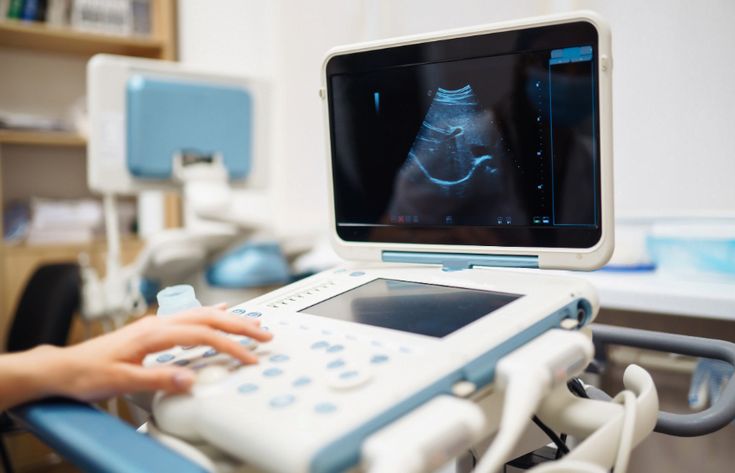
After obtaining the Doppler spectrum, the operator may need to identify or trace the spectral envelope to calculate parameters such as PSV (Peak Systolic Velocity), EDV (End-Diastolic Velocity), RI (Resistance Index), PI (Pulsatility Index), and the S/D ratio.
This process has several limitations:
Automation and AI-based tools are therefore being developed to support recognition, measurement, and standardization of certain steps in the workflow, helping physicians reduce repetitive tasks and focus more on clinical interpretation.
Doppler ultrasound diagnostic support software refers to software tools that apply AI and advanced medical image-processing algorithms and are either integrated directly into ultrasound systems or operated through PACS/EMR infrastructure.
Instead of requiring physicians to perform measurements and manually trace spectral waveforms, the software can automatically recognize vascular or cardiac structures, analyze waveform patterns, and instantly extract important hemodynamic parameters:
Depending on the system configuration and software, modern ultrasound systems may provide multiple Doppler modes for different clinical applications.

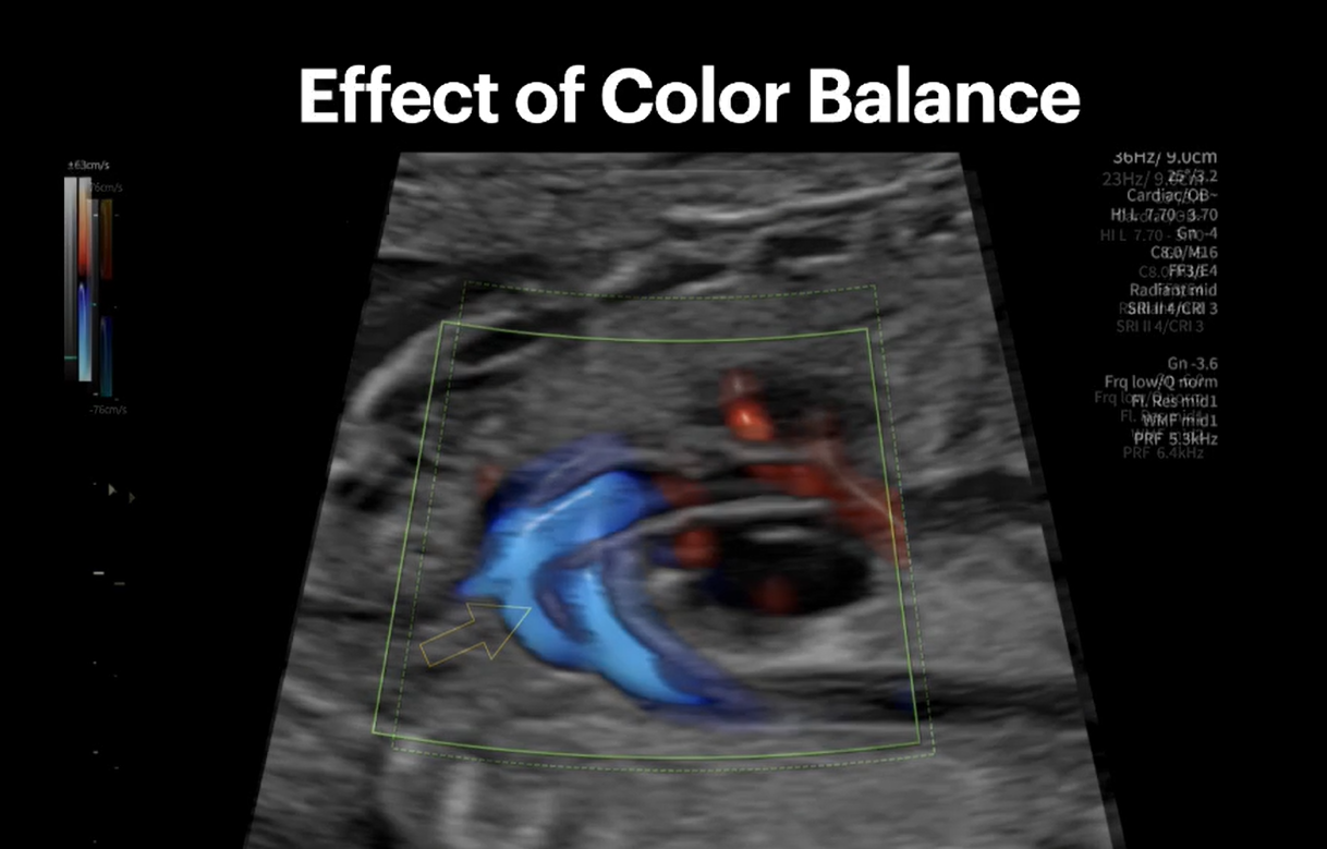
Color Doppler uses color coding to display blood flow information on a 2D ultrasound image, allowing physicians to quickly observe the direction and characteristics of blood flow within the region of interest. This technique is commonly used to identify blood vessels and detect areas of abnormal flow.
Pulsed Wave (PW) Doppler enables blood flow sampling at a specific location and displays a velocity waveform over time. It is an important mode for measuring parameters such as PSV, EDV, RI, PI, and the S/D ratio in vascular and obstetric examinations.
Continuous Wave (CW) Doppler can record high-velocity blood flow and is particularly useful in echocardiography, such as when assessing flow across heart valves or in cases where blood flow velocity exceeds the measurement limits of PW Doppler.
Power Doppler displays the strength of the Doppler signal rather than primarily representing flow direction. This technique offers good sensitivity to low-velocity blood flow and can support the assessment of perfusion in small vessels or areas with weak blood flow signals.
Tissue Doppler Imaging (TDI) is primarily used in echocardiography to measure myocardial motion velocity, supporting the assessment of systolic and diastolic cardiac function.
Integrating Doppler modes with automated measurement tools, image processing, and intelligent algorithms helps modern ultrasound systems optimize examination workflows. However, specific automation or AI capabilities vary depending on the ultrasound system, software version, and installed application package.
The software can automatically delineate spectral waveforms and rapidly calculate blood flow parameters from Doppler examinations:
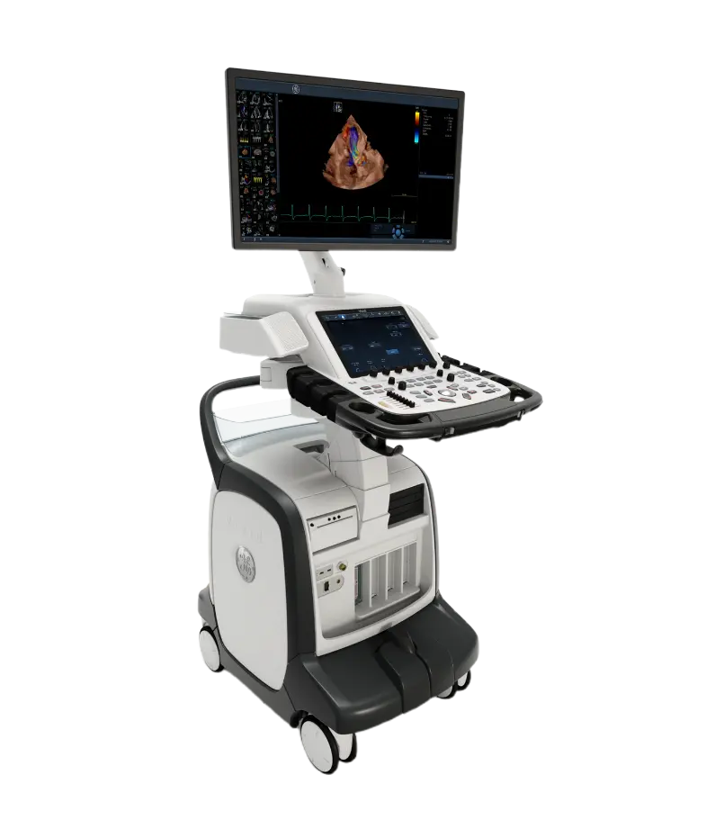
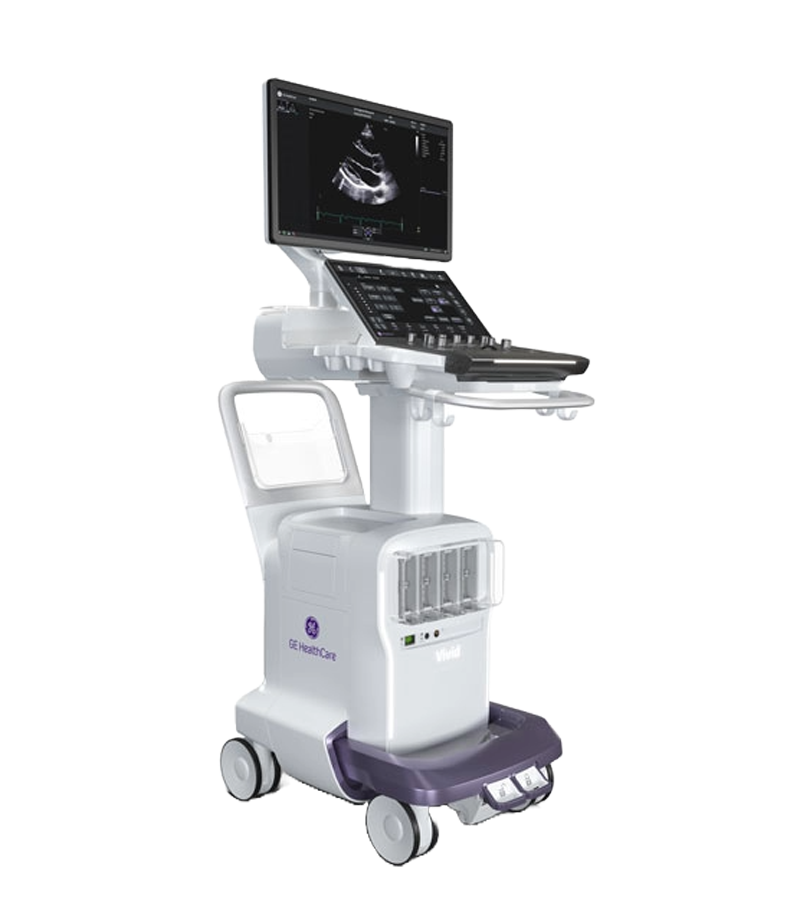
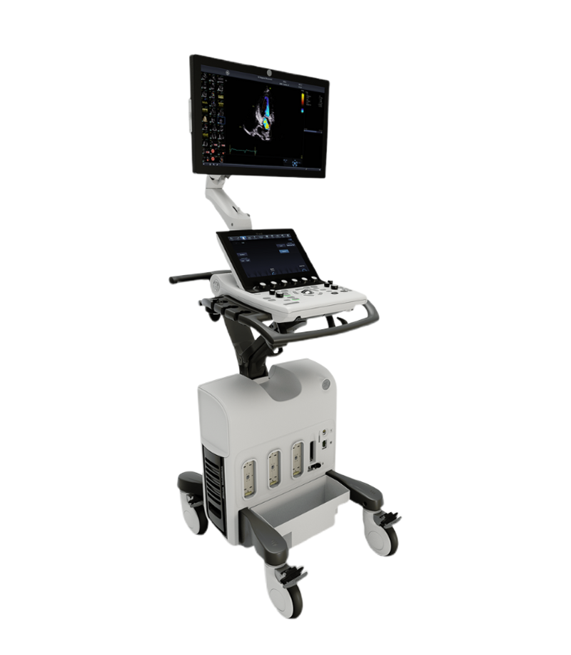
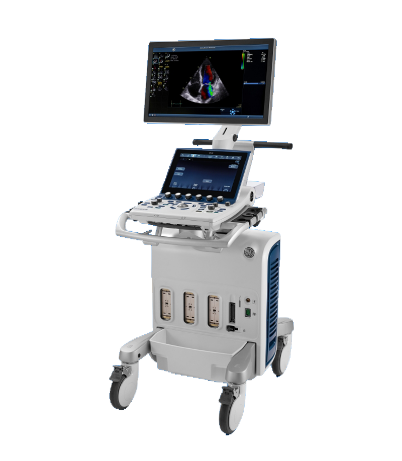
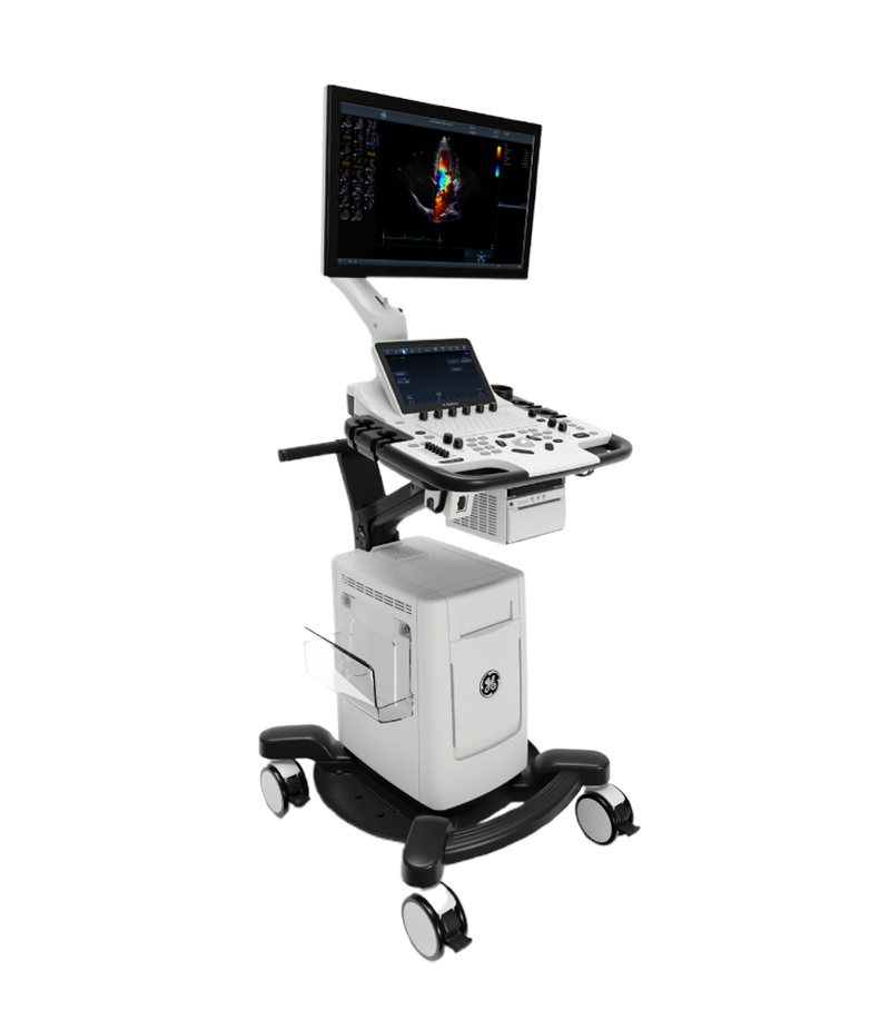
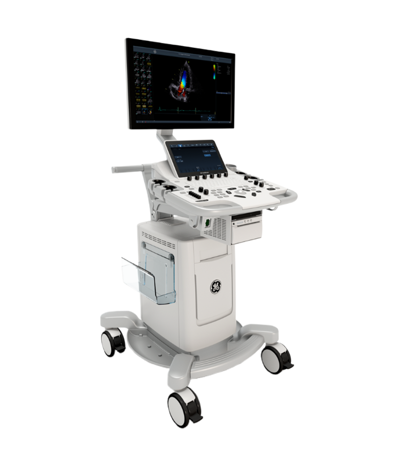
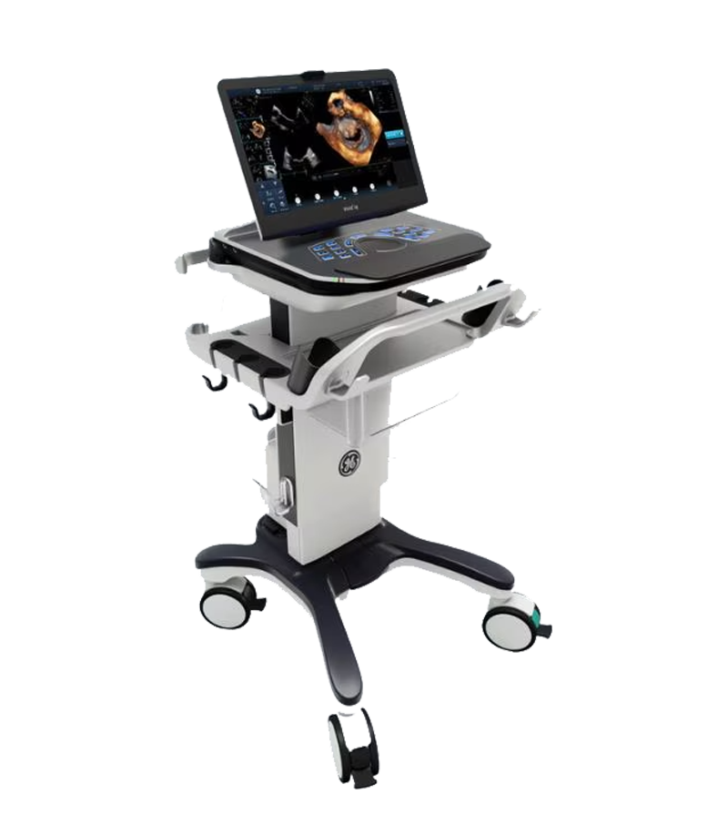
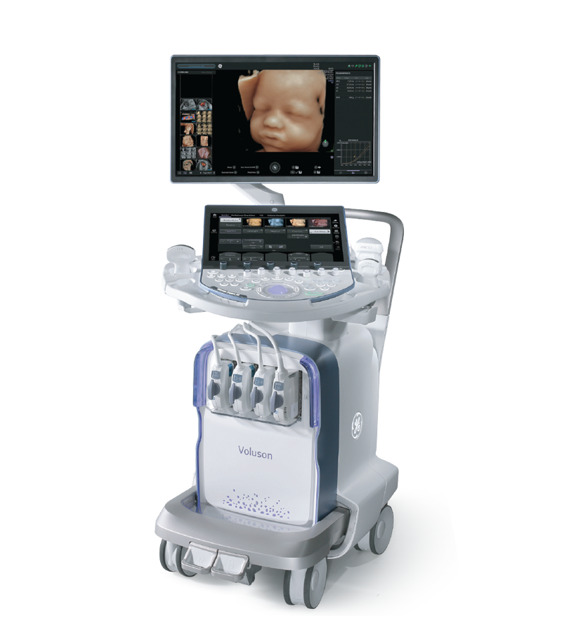
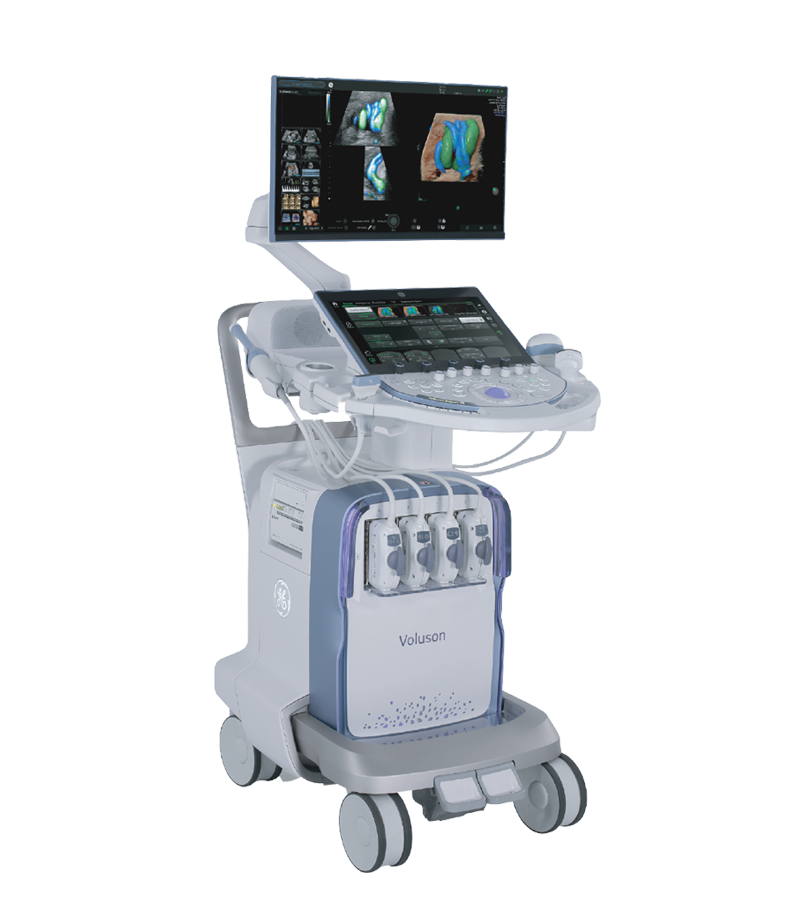
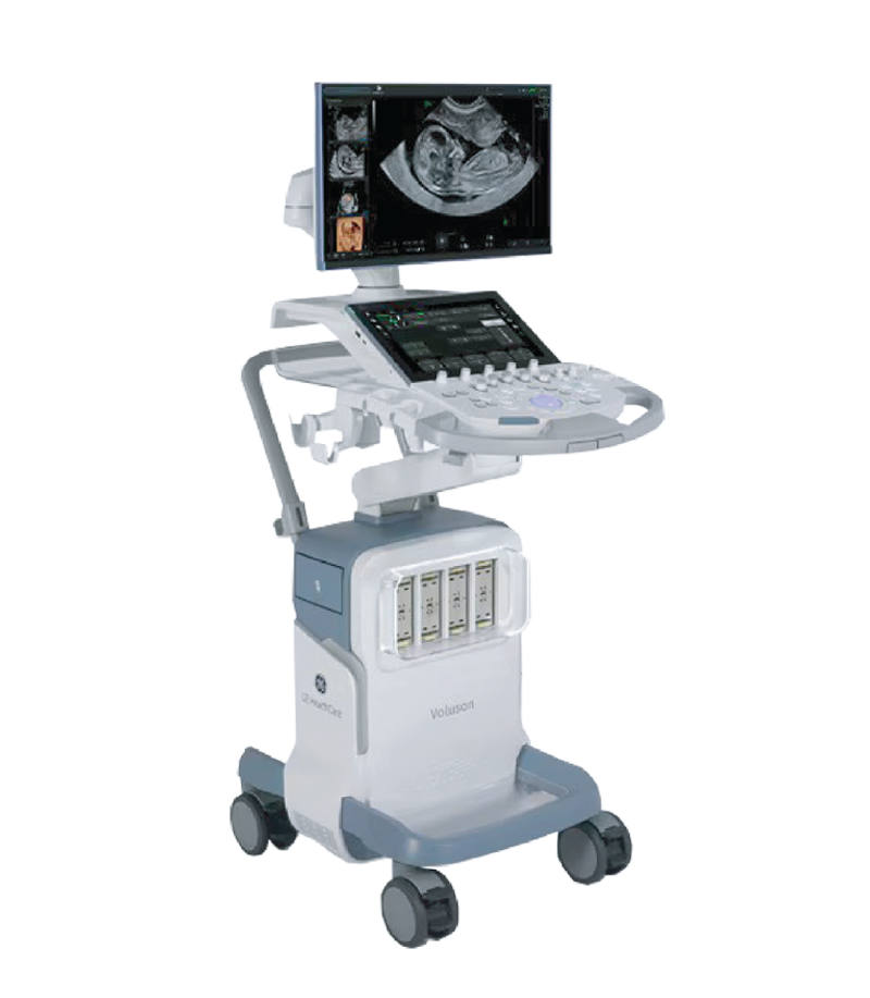
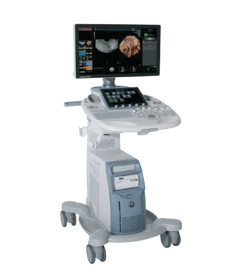
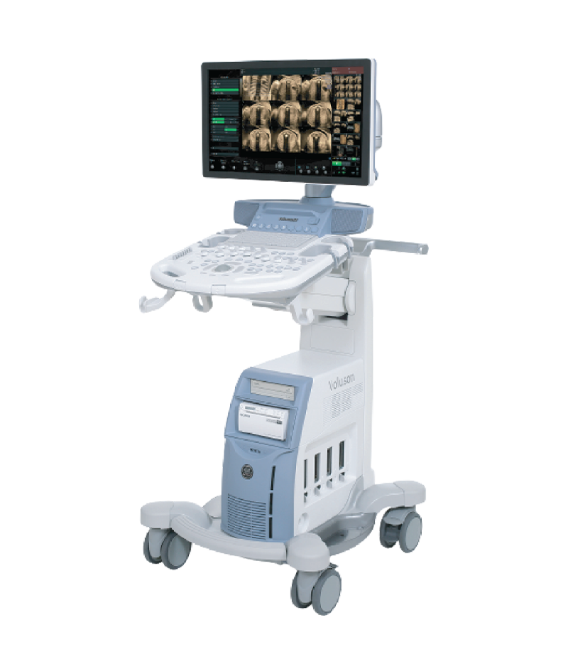
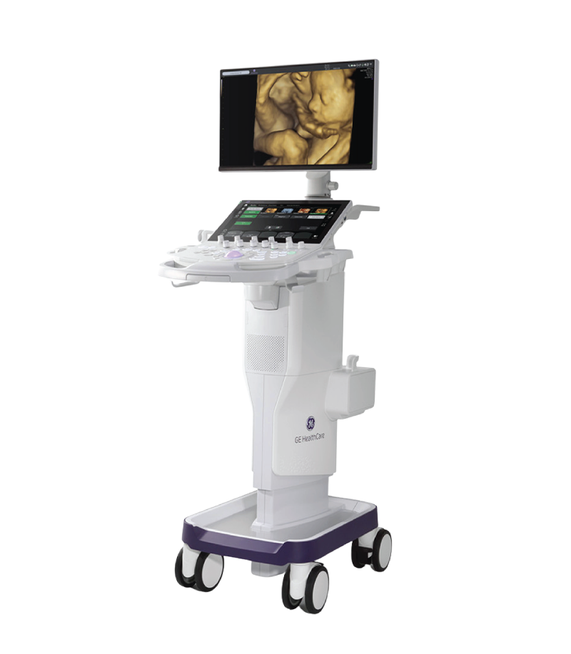
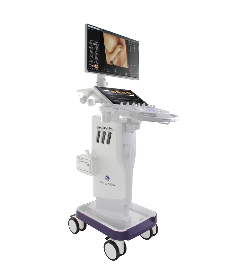
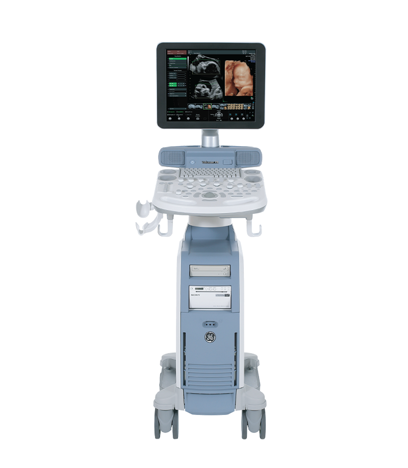
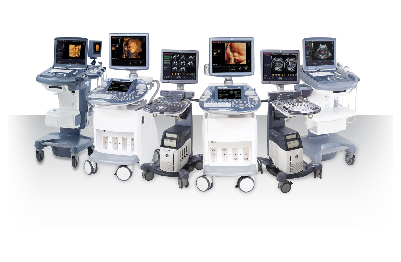
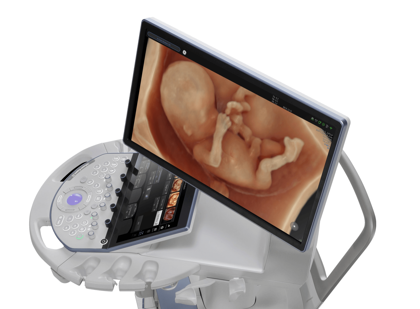
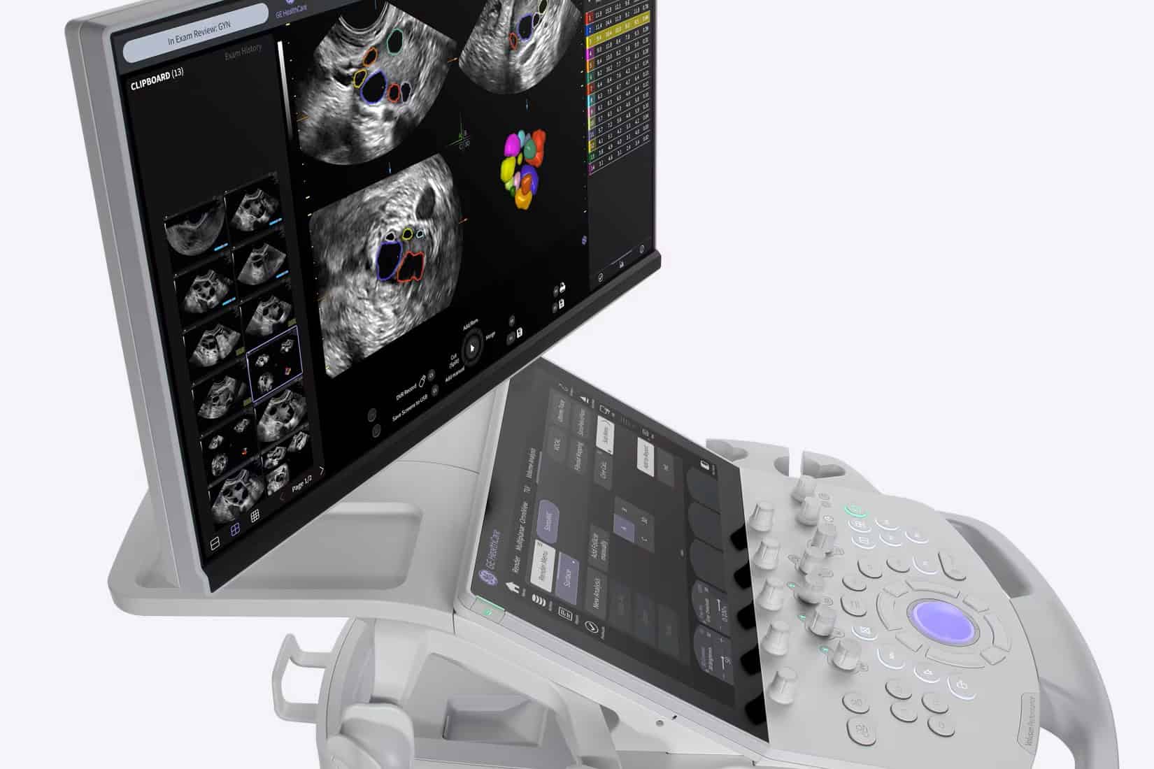
The reliability of AI software depends significantly on the quality of data provided by the ultrasound hardware. TD TECH is proud to provide leading obstetric, gynecologic, and cardiovascular ultrasound solutions from GE HealthCare, featuring advanced intelligent Doppler technologies:
Cardiac and Obstetric Ultrasound Systems
The application of AI-integrated Doppler ultrasound diagnostic support software represents a strategic step for healthcare facilities seeking to standardize clinical quality, reduce diagnostic variability, and enhance clinical performance.
Ultrasound is a core tool in pregnancy care, from confirming early pregnancy and monitoring fetal growth to assessing fetal anatomy, the fetal heart, and Doppler flow. Therefore, when choosing an obstetric ultrasound system, image quality...
Xem thêm
GE HealthCare is a global medical technology company operating across diagnostic imaging, ultrasound, patient monitoring, diagnostic pharmaceuticals, AI, and healthcare software solutions. GE HealthCare offers a broad portfolio of ultrasound systems, with each product family...
Xem thêm
Congenital heart disease is among the most common congenital abnormalities in fetuses; however, prenatal detection remains challenging in routine obstetric practice. Although professional organizations such as ISUOG have issued detailed guidelines on the fetal cardiac...
Xem thêm
What Is 2D Ultrasound? 2D ultrasound is a technique that produces real-time, two-dimensional cross-sectional images of structures inside the body. The transducer emits ultrasound waves into the body and receives reflected signals from tissues. The...
Xem thêm
What Is Twin-to-Twin Transfusion Syndrome? Twin-to-Twin Transfusion Syndrome (TTTS) is a serious complication that can occur in monochorionic twin pregnancies, in which vascular anastomoses within the shared placenta lead to an imbalanced exchange of blood...
Xem thêm
In obstetrics and gynecology, the growing need for clearer fetal visualization and more efficient examination workflows is driving the development of next-generation 3D/4D ultrasound systems. These devices are often searched for under the term “5D...
Xem thêm



 Xem video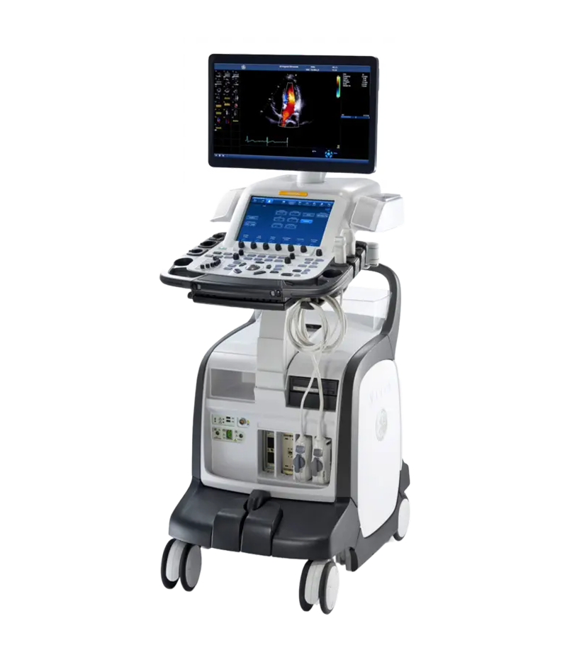




 Consult Now