 Email: tdmedical@gmail.com


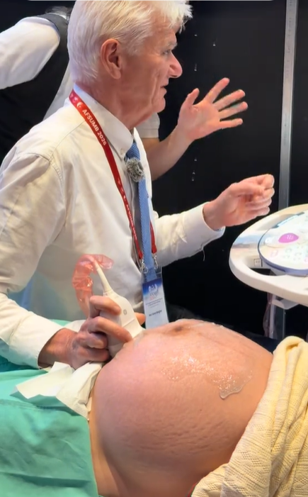
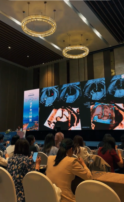
Professor Simon Meagher and Professor Ritsuko Pooh Performing Ultrasound Examinations Using the Voluson E22 System
 8.3K
8.3K
 51
51
 03-Sep-2026
 89,775
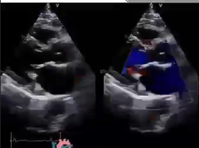
The same valve, the same ultrasound screen—yet they can tell three completely different stories.
Video source: Dr. Shubham
This video illustrates the appearance of the mitral valve in three different conditions: a normal mitral valve, rheumatic heart disease (RHD), and mitral valve calcification.
At first glance, the images may appear quite similar. However, from a clinical perspective, each condition has its own distinctive features and carries different diagnostic implications.
The valve leaflets are thin, pliable, and move freely during opening and closure. Blood flow across the valve is typically smooth and relatively uniform on color Doppler imaging. This reflects a valve that is functioning efficiently and working in coordination with the heart.
The valve leaflets may become thickened, particularly at the tips, with restricted mobility and a characteristic appearance often described as “hockey-stick” deformity. Blood flow across the valve may become turbulent as a result of mitral stenosis, regurgitation, or a combination of both.
These changes can develop as long-term consequences of rheumatic heart disease and may affect valve function for many years.
On ultrasound, calcified areas typically appear highly echogenic, particularly around the mitral annulus. Calcification reduces the flexibility of the valve apparatus and may alter blood flow across the valve.
This is commonly associated with degenerative changes over time and may also be related to various metabolic and cardiovascular factors.
Rheumatic valve disease primarily causes thickening, fibrosis, and scarring of the valve leaflets and associated structures. Mitral annular calcification alters the structure and flexibility of the valve annulus. In contrast, a normal mitral valve remains thin, flexible, and mobile, allowing blood to flow efficiently.
Distinguishing between these conditions is important for disease assessment, prognosis, and determining the most appropriate approach to follow-up or treatment.
Echocardiography is not simply about producing images. It combines structural recognition, blood-flow assessment, and clinical reasoning.
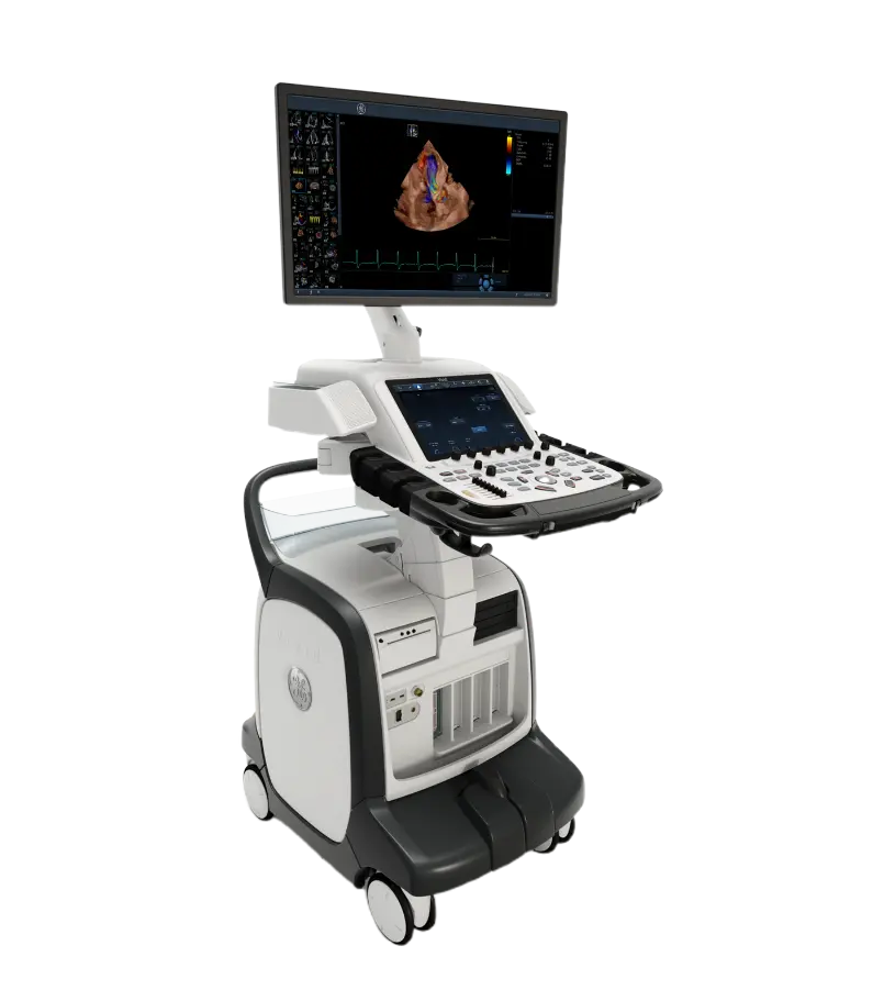
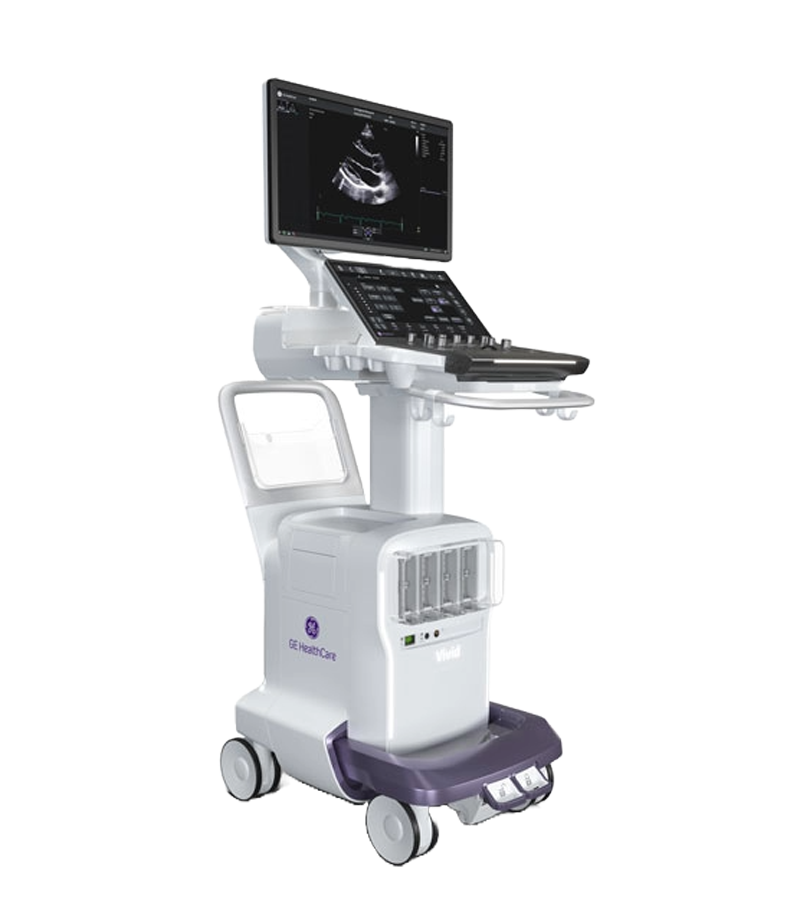
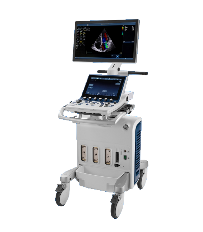
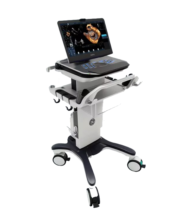
Vivid Echocardiography System
The images were acquired using the Vivid cardiovascular and general imaging ultrasound system by GE HealthCare.
Video source: Dr. Shubham
The emergence of diagnostic support software for Doppler ultrasound integrated with artificial intelligence (AI) has marked a major breakthrough, helping automate measurements, improve accuracy, and optimize workflow efficiency for physicians. What Is Doppler Ultrasound and...
Xem thêm
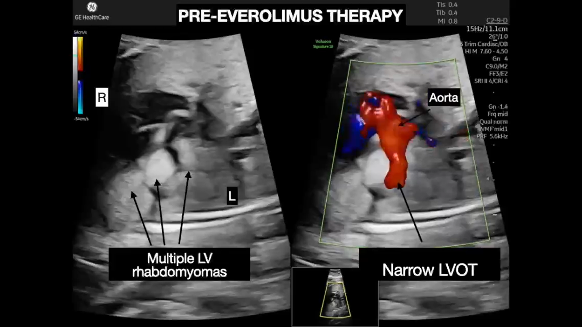
At 24 weeks of pregnancy, a routine screening ultrasound revealed multiple masses within the fetal heart. A subsequent fetal echocardiogram confirmed the presence of rhabdomyomas involving both ventricles. Although cardiac rhabdomyomas are generally benign...
Xem thêm
Congenital heart disease is among the most common congenital abnormalities in fetuses; however, prenatal detection remains challenging in routine obstetric practice. Although professional organizations such as ISUOG have issued detailed guidelines on the fetal cardiac...
Xem thêm
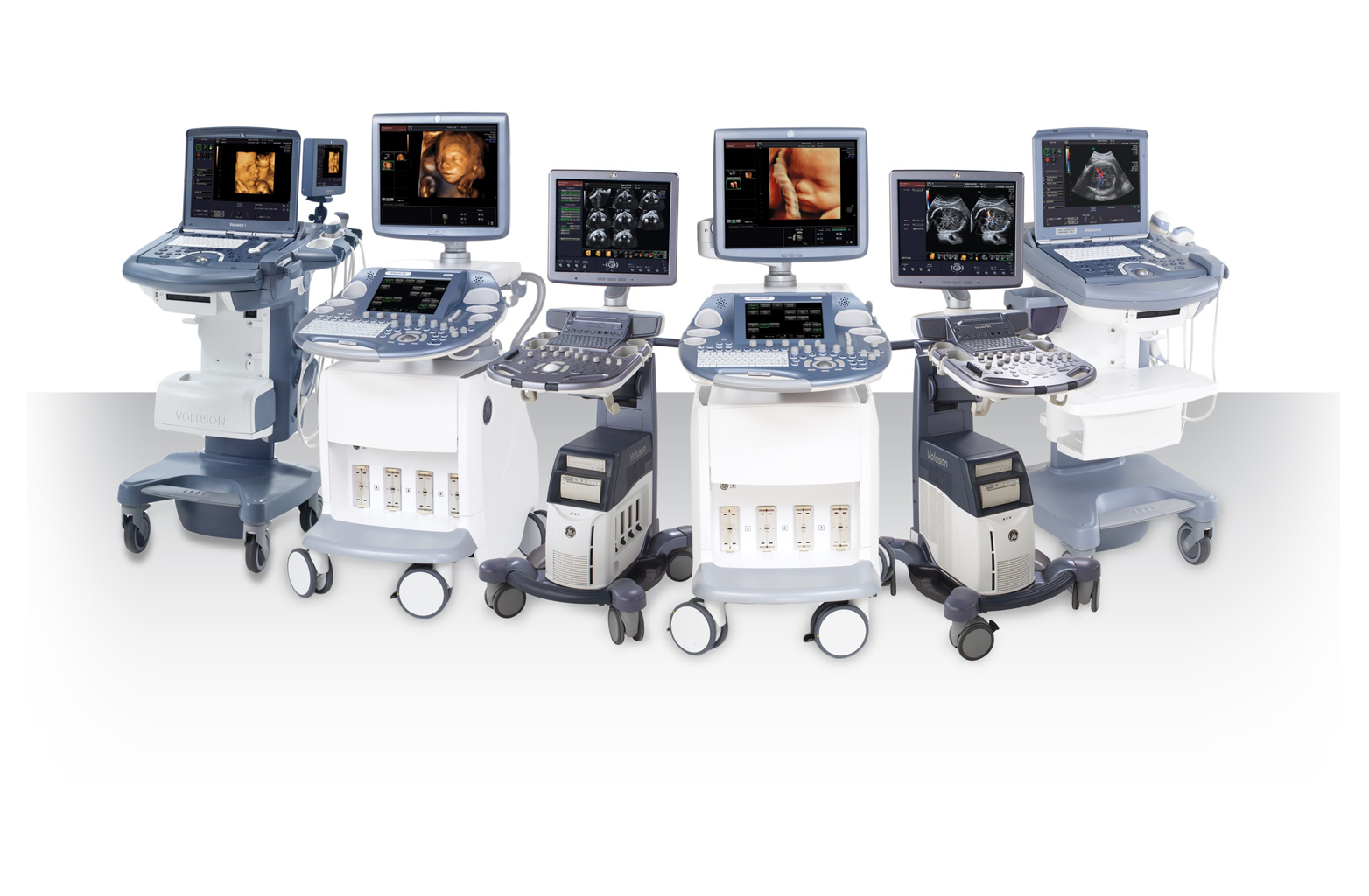
Ultrasound is a core tool in pregnancy care, from confirming early pregnancy and monitoring fetal growth to assessing fetal anatomy, the fetal heart, and Doppler flow. Therefore, when choosing an obstetric ultrasound system, image quality...
Xem thêm
Unboxing the Voluson P16 and P18 ultrasound machines – the two latest models in the P-Series featuring modern designs, sharp imagery, and effective support for OB/GYN and multi-specialty diagnostics. Introduction In the field of diagnostic...
Xem thêm
What Is Twin-to-Twin Transfusion Syndrome? Twin-to-Twin Transfusion Syndrome (TTTS) is a serious complication that can occur in monochorionic twin pregnancies, in which vascular anastomoses within the shared placenta lead to an imbalanced exchange of blood...
Xem thêm


 Xem video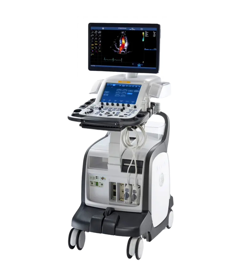




 Consult Now